

Alkaline Phosphatase (Placental) / PLAP (Germ Cell Tumor Marker)(ALPP/516), Biotin conjugate, 0.1mg/mL [26628-22-8]
BNCB0516
ReactivityBovine, Human, Mouse
Product group Antibodies
TargetALPP
Overview
- SupplierBiotium
- Product NameAlkaline Phosphatase (Placental) / PLAP (Germ Cell Tumor Marker)(ALPP/516), Biotin conjugate, 0.1mg/mL [26628-22-8]
- Delivery Days Customer9
- CAS Number26628-22-8
- CertificationResearch Use Only
- ClonalityMonoclonal
- Clone IDALPP/516
- Concentration0.1 mg/ml
- ConjugateBiotin
- Gene ID250
- Target nameALPP
- Target descriptionalkaline phosphatase, placental
- Target synonymsALP, ALPI, IAP, PALP, PLAP, PLAP-1, alkaline phosphatase, placental type, Intestinal alkaline phosphatase, Intestinal-type alkaline phosphatase, alkaline phosphatase Regan isozyme, alkaline phosphomonoesterase, glycerophosphatase, placental alkaline phosphatase 1
- HostMouse
- IsotypeIgG1
- Protein IDP05187
- Protein NameAlkaline phosphatase, placental type
- Scientific DescriptionThis antibody reacts with placental alkaline phosphatase. There are at least four distinct but related alkaline phosphatases: intestinal, placental (PLAP), placental-like, and liver/bone/kidney (tissue non-specific). The first three are located together on chromosome 2, while the tissue non-specific form is located on chromosome 1. PLAP is a tissue specific, trophoblast-derived, 70 kDa, glycosyl-phosphatidylinositol (GPI)-anchored, dimeric, zinc metallo-glycoprotein that catalyzes the hydrolysis of phosphomonoesters into an inorganic phosphate and an alcohol. It is present in the placenta and serum of pregnant women and in high frequency in gynecological and testicular cancers and in lower frequency in other tumors. The three tissue-specific APs in humans, PLAP, germ cell AP (GCAP) and intestinal AP, are 90-98% homologous. Non-tissue specific AP is found in kidney, liver and bone. This MAb binds equally well to all common allelic variants (S, F, FS and I) of PLAP and to some variants of AP from normal human testis. This MAb can be used as tracer antibody in ELISA to detect PLAP in serum of S, F, FS and I phenotypes. Primary antibodies are available purified, or with a selection of fluorescent CF® Dyes and other labels. CF® Dyes offer exceptional brightness and photostability. Note: Conjugates of blue fluorescent dyes like CF®405S and CF®405M are not recommended for detecting low abundance targets, because blue dyes have lower fluorescence and can give higher non-specific background than other dye colors.
- SourceAnimal
- ReactivityBovine, Human, Mouse
- Storage Instruction2°C to 8°C,RT
- UNSPSC41116161
MSDS
Related products
Product group Antibodies
Anti-ALPP AntibodyA29021
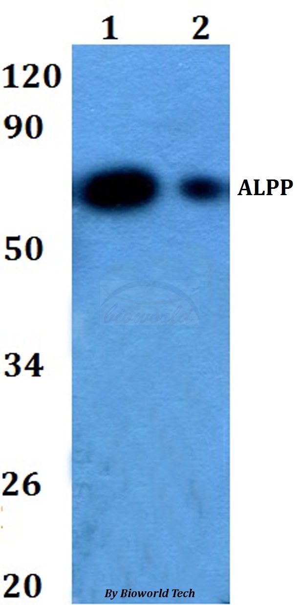
ApplicationsWestern Blot
ReactivityHuman, Mouse
- SizePrice
Product group Antibodies

Anti-PLAP [10G6D9E8]Ab03404-1.1
ApplicationsWestern Blot, ImmunoHistoChemistry
ReactivityHuman
TargetALPP
- SizePrice

Product group Antibodies
Anti-Placental alkaline phosphatase (PLAP)/ALPP Antibody Picoband(r)A01718-CARRIER-FREE
ApplicationsFlow Cytometry, ImmunoFluorescence, Western Blot, ImmunoCytoChemistry, ImmunoHistoChemistry
ReactivityHuman
TargetALPP
- SizePrice
Product group Antibodies
ApplicationsFlow Cytometry, Western Blot, ELISA
ReactivityHuman, Mouse
TargetALPP
- SizePrice
Product group Antibodies
ALPP AntibodyCSB-PA001632ESR1HU
ApplicationsWestern Blot, ELISA, ImmunoHistoChemistry
ReactivityHuman
TargetALPP
- SizePrice
Product group Antibodies
Alpp Polyclonal AntibodyCAC10640
ApplicationsWestern Blot, ELISA, ImmunoHistoChemistry
TargetALPP
- SizePrice
![IHC-P analysis of human placenta (mature) tissue using GTX04413 Placental Alkaline Phosphatase antibody [MSVA-350R] HistoMAX?. Placental Alkaline Phosphatase immunostaining increases continuously throughout the pregnancy. Placental Alkaline Phosphatase immunostaining is strong in the cyto- and syncytiotrophoblast of the mature placenta.](https://www.genetex.com/upload/website/prouct_img/normal/GTX04413/GTX04413_20230728_IHC-P_98_23072722_473.webp)
Product group Antibodies
ApplicationsImmunoHistoChemistry, ImmunoHistoChemistry Paraffin
ReactivityHuman
TargetALPP
- SizePrice
Product group Antibodies

ApplicationsWestern Blot, ELISA
ReactivityHuman
TargetALPP
- SizePrice