

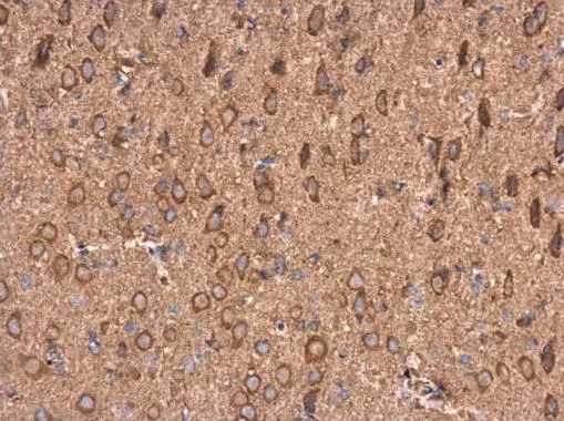
AMPD2 antibody [N3C3] detects AMPD2 protein at cytoplasm in rat brain by immunohistochemical analysis. Sample: Paraffin-embedded rat brain. AMPD2 antibody [N3C3] (GTX112483) diluted at 1:500.
Antigen Retrieval: Citrate buffer, pH 6.0, 15 min
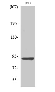
![Various whole cell extracts (30 μg) were separated by 7.5% SDS-PAGE, and the membrane was blotted with AMPD2 antibody [N3C3] (GTX112483) diluted at 1:1000.](https://www.genetex.com/upload/website/prouct_img/normal/GTX112483/GTX112483_40150_20160428_WB_w_23060500_371.webp "Various whole cell extracts (30 μg) were separated by 7.5% SDS-PAGE, and the membrane was blotted with AMPD2 antibody [N3C3] (GTX112483) diluted at 1:1000.")
![AMPD2 antibody [N3C3] detects AMPD2 protein by immunohistochemical analysis. Sample: Frozen-sectioned rat E13.5 brain. Green: AMPD2 stained by AMPD2 antibody [N3C3] (GTX112483) diluted at 1:250. Red: beta Tubulin 3/ Tuj1, stained by beta Tubulin 3/ Tuj1 antibody [GT1338] (GTX631831) diluted at 1:500. Blue: Fluoroshield with DAPI (GTX30920).](https://www.genetex.com/upload/website/prouct_img/normal/GTX112483/GTX112483_40150_20171106_IHC-Fr_R_w_23060500_975.webp "AMPD2 antibody [N3C3] detects AMPD2 protein by immunohistochemical analysis. Sample: Frozen-sectioned rat E13.5 brain. Green: AMPD2 stained by AMPD2 antibody [N3C3] (GTX112483) diluted at 1:250. Red: beta Tubulin 3/ Tuj1, stained by beta Tubulin 3/ Tuj1 antibody [GT1338] (GTX631831) diluted at 1:500. Blue: Fluoroshield with DAPI (GTX30920).")
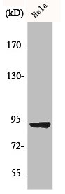
 A: Hela 7.5% SDS PAGE GTX112483 diluted at 1:1000")
AMPD2 antibody [N3C3] detects AMPD2 protein at cytoplasm in rat brain by immunohistochemical analysis. Sample: Paraffin-embedded rat brain. AMPD2 antibody [N3C3] (GTX112483) diluted at 1:500.
Antigen Retrieval: Citrate buffer, pH 6.0, 15 min
AMPD2 antibody [N3C3]
GTX112483
ApplicationsWestern Blot, ImmunoHistoChemistry, ImmunoHistoChemistry Frozen, ImmunoHistoChemistry Paraffin
Product group Antibodies
ReactivityHuman, Rat
TargetAMPD2
Overview
- SupplierGeneTex
- Product NameAMPD2 antibody [N3C3]
- Delivery Days Customer9
- Application Supplier NoteWB: 1:500-1:3000. IHC-P: 1:100-1:1000. IHC-Fr: 1:100-1:1000. *Optimal dilutions/concentrations should be determined by the researcher.Not tested in other applications.
- ApplicationsWestern Blot, ImmunoHistoChemistry, ImmunoHistoChemistry Frozen, ImmunoHistoChemistry Paraffin
- CertificationResearch Use Only
- ClonalityPolyclonal
- Concentration0.99 mg/ml
- ConjugateUnconjugated
- Gene ID271
- Target nameAMPD2
- Target descriptionadenosine monophosphate deaminase 2
- Target synonymsAMPD, PCH9, SPG63, AMP deaminase 2, adenosine monophosphate deaminase 2 (isoform L)
- HostRabbit
- IsotypeIgG
- Protein IDQ01433
- Protein NameAMP deaminase 2
- Scientific DescriptionAdenosine monophosphate deaminase-2 (EC 3.5.4.6) catalyzes the deamination of AMP to IMP and plays an important role in the purine nucleotide cycle.[supplied by OMIM]
- ReactivityHuman, Rat
- Storage Instruction-20°C or -80°C,2°C to 8°C
- UNSPSC41116161
Datasheet
Related products
Product group Antibodies
Anti-AMPD2 AntibodyA97718
ApplicationsWestern Blot, ELISA
ReactivityHuman, Mouse, Rat
- SizePrice
Product group Antibodies
Anti-AMPD2 (Center) Antibody102-27662
ApplicationsFlow Cytometry, Western Blot, ImmunoHistoChemistry, ImmunoHistoChemistry Paraffin
TargetAMPD2
- SizePrice
Product group Antibodies
AMPD2 AntibodyCSB-PA000869
ApplicationsWestern Blot, ELISA
ReactivityHuman, Mouse, Rat
TargetAMPD2
- SizePrice
Product group Antibodies
AMPD2 Polyclonal AntibodyCAC14704
ApplicationsImmunoFluorescence, Western Blot, ELISA, ImmunoHistoChemistry
TargetAMPD2
- SizePrice
Product group Antibodies

AMPD2 Antibody (Biotin)LS-C498371
ApplicationsELISA
ReactivityHuman
TargetAMPD2
- SizePrice

Product group Antibodies
Anti-AMPD2 AntibodyHPA045760
ApplicationsWestern Blot, ImmunoCytoChemistry, ImmunoHistoChemistry
ReactivityHuman
TargetAMPD2
- SizePrice