


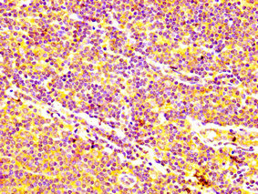
Immunohistochemical staining of human kidney shows strong membranous positivity in cells in tubules.
Immunohistochemical staining of human kidney shows strong membranous positivity in cells in tubules.
Anti-ABCC5 Antibody
HPA052295
ApplicationsImmunoHistoChemistry
Product group Antibodies
ReactivityHuman
TargetABCC5
Overview
- SupplierAtlas Antibodies
- Product NameAnti-ABCC5 Antibody
- Delivery Days Customer4
- ApplicationsImmunoHistoChemistry
- CertificationResearch Use Only
- ClonalityPolyclonal
- ConjugateUnconjugated
- Gene ID10057
- Target nameABCC5
- Target descriptionATP binding cassette subfamily C member 5
- Target synonymsABC33, EST277145, MOAT-C, MOATC, MRP5, SMRP, pABC11, ATP-binding cassette sub-family C member 5, ATP-binding cassette, sub-family C (CFTR/MRP), member 5, canalicular multispecific organic anion transporter C, multi-specific organic anion transporter C, multidrug resistance-associated protein 5
- HostRabbit
- IsotypeIgG
- Protein IDO15440
- Protein NameATP-binding cassette sub-family C member 5
- Scientific DescriptionRecombinant Protein Epitope Signature Tag (PrEST) antigen sequence
- ReactivityHuman
- Storage Instruction-20°C,2°C to 8°C
- UNSPSC41116161
Datasheet
MSDS
Related products
Product group Antibodies
ABCC5 AntibodyCSB-PA001064LA01HU
ApplicationsImmunoFluorescence, ELISA, ImmunoHistoChemistry
ReactivityHuman
TargetABCC5
- SizePrice
Product group Antibodies
Anti-MRP5 AntibodyA14310
ApplicationsWestern Blot
ReactivityHuman, Mouse, Rat
- SizePrice
Product group Antibodies
Anti-ABCC5 Antibody144-65635
ApplicationsWestern Blot
ReactivityHuman, Mouse, Rat
TargetABCC5
- SizePrice

Product group Antibodies
Anti-MRP5/ABCC5 AntibodyA03040-1-CARRIER-FREE
ApplicationsFlow Cytometry, ELISA, ImmunoHistoChemistry
ReactivityHuman
TargetABCC5
- SizePrice
Product group Antibodies

Goat anti-MRP5EB08699
ApplicationsELISA, ImmunoHistoChemistry
ReactivityBovine, Canine, Human, Mouse, Porcine, Rat
TargetABCC5
- SizePrice

Product group Antibodies
Anti-ABCC5 AntibodyHPA044067
ApplicationsImmunoCytoChemistry
ReactivityHuman
TargetABCC5
- SizePrice
Product group Antibodies
Anti-ABCC5 AntibodyHPA044067
ApplicationsImmunoCytoChemistry
ReactivityHuman
TargetABCC5
- SizePrice
Product group Antibodies

ABCC5 / MRP5 AntibodyLS-C402447
ApplicationsWestern Blot, ELISA, ImmunoHistoChemistry
ReactivityHuman, Mouse, Rat
TargetABCC5
- SizePrice