


Immunohistochemical staining of human skin shows moderate to strong membranous positivity in squamous epithelial cells.
Immunohistochemical staining of human skin shows moderate to strong membranous positivity in squamous epithelial cells.
Anti-ACVR2A Antibody
HPA046997
ApplicationsImmunoHistoChemistry
Product group Antibodies
ReactivityHuman
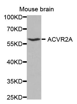
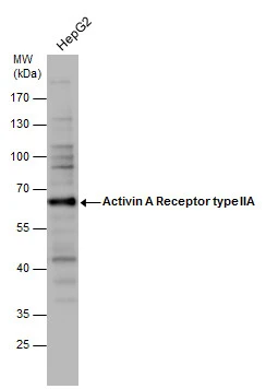
TargetACVR2A
Overview
- SupplierAtlas Antibodies
- Product NameAnti-ACVR2A Antibody
- Delivery Days Customer4
- ApplicationsImmunoHistoChemistry
- CertificationResearch Use Only
- ClonalityPolyclonal
- ConjugateUnconjugated
- Gene ID92
- Target nameACVR2A
- Target descriptionactivin A receptor type 2A
- Target synonymsACTRII, ACVR2, activin receptor type-2A, activin A receptor, type IIA
- HostRabbit
- IsotypeIgG
- Protein IDP27037
- Protein NameActivin receptor type-2A
- Scientific DescriptionRecombinant Protein Epitope Signature Tag (PrEST) antigen sequence
- ReactivityHuman
- Storage Instruction-20°C,2°C to 8°C
- UNSPSC41116161
Datasheet
MSDS
Related products

Product group Antibodies
Anti-ACVR2A Antibody Picoband(r)A04770-2-CARRIER-FREE
ApplicationsWestern Blot, ELISA
ReactivityHuman
TargetACVR2A
- SizePrice
Product group Antibodies
Anti-ACVR2A Antibody144-01981
ApplicationsImmunoFluorescence, Western Blot, ImmunoHistoChemistry
ReactivityHuman, Mouse, Rat
TargetACVR2A
- SizePrice
Product group Antibodies
Anti-ACVR2A AntibodyA42474
ApplicationsWestern Blot
ReactivityHuman, Mouse, Rat
- SizePrice
Product group Antibodies
ACVR2A Polyclonal AntibodyBS-22941R
ApplicationsWestern Blot
ReactivityHuman, Mouse, Rat
TargetACVR2A
- SizePrice

Product group Antibodies
ACVR2A AntibodyCSB-PA001260ESR1HU
ApplicationsELISA, ImmunoHistoChemistry
ReactivityHuman
TargetACVR2A
- SizePrice
Product group Antibodies
ApplicationsImmunoPrecipitation, Western Blot, ImmunoCytoChemistry, ImmunoHistoChemistry
ReactivityMouse, Rat
TargetACVR2A
- SizePrice
Product group Antibodies

ACVR2 / ACVR2A AntibodyLS-C402149
ApplicationsWestern Blot, ELISA, ImmunoHistoChemistry
ReactivityHuman, Mouse, Rat
TargetACVR2A
- SizePrice
Product group Antibodies
ApplicationsWestern Blot
ReactivityHuman
TargetACVR2A
- SizePrice