


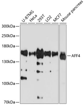
Figure 1. Western blot analysis of AFF4 using anti-AFF4 antibody (A03824). Electrophoresis was performed on a 5-20% SDS-PAGE gel at 70V (Stacking gel) / 90V (Resolving gel) for 2-3 hours. The sample well of each lane was loaded with 30 ug of sample under reducing conditions. Lane 1: human Hela whole cell lysates, Lane 2: human HepG2 whole cell lysates, Lane 3: human 293T whole cell lysates, Lane 4: human U87 whole cell lysates. After electrophoresis, proteins were transferred to a nitrocellulose membrane at 150 mA for 50-90 minutes. Blocked the membrane with 5% non-fat milk/TBS for 1.5 hour at RT. The membrane was incubated with rabbit anti-AFF4 antigen affinity purified polyclonal antibody (Catalog # A03824) at 0.5 microg/mL overnight at 4°C, then washed with TBS-0.1%Tween 3 times with 5 minutes each and probed with a goat anti-rabbit IgG-HRP secondary antibody at a dilution of 1:5000 for 1.5 hour at RT. The signal is developed using an Enhanced Chemiluminescent detection (ECL) kit (Catalog # EK1002) with Tanon 5200 system. A specific band was detected for AFF4 at approximately 150 kDa. The expected band size for AFF4 is at 127 kDa.
. Overlay histogram showing SiHa cells stained with A03824 (Blue line). To facilitate intracellular staining, cells were fixed with 4% paraformaldehyde and permeabilized with permeabilization buffer. The cells were blocked with 10% normal goat serum. And then incubated with rabbit anti-AFF4 Antibody (A03824,1microg/1x106 cells) for 30 min at 20°C. DyLight®488 conjugated goat anti-rabbit IgG (BA1127, 5-10microg/1x106 cells) was used as secondary antibody for 30 minutes at 20°C. Isotype control antibody (Green line) was rabbit IgG (1microg/1x106) used under the same conditions. Unlabelled sample without incubation with primary antibody and secondary antibody (Red line) was used as a blank control.")
. AFF4 was detected in a paraffin-embedded section of human placenta tissue. Heat mediated antigen retrieval was performed in EDTA buffer (pH 8.0, epitope retrieval solution). The tissue section was blocked with 10% goat serum. The tissue section was then incubated with 2 microg/ml rabbit anti-AFF4 Antibody (A03824) overnight at 4°C. Peroxidase Conjugated Goat Anti-rabbit IgG was used as secondary antibody and incubated for 30 minutes at 37°C. The tissue section was developed using HRP Conjugated Rabbit IgG Super Vision Assay Kit (Catalog # SV0002) with DAB as the chromogen.")
Figure 1. Western blot analysis of AFF4 using anti-AFF4 antibody (A03824). Electrophoresis was performed on a 5-20% SDS-PAGE gel at 70V (Stacking gel) / 90V (Resolving gel) for 2-3 hours. The sample well of each lane was loaded with 30 ug of sample under reducing conditions. Lane 1: human Hela whole cell lysates, Lane 2: human HepG2 whole cell lysates, Lane 3: human 293T whole cell lysates, Lane 4: human U87 whole cell lysates. After electrophoresis, proteins were transferred to a nitrocellulose membrane at 150 mA for 50-90 minutes. Blocked the membrane with 5% non-fat milk/TBS for 1.5 hour at RT. The membrane was incubated with rabbit anti-AFF4 antigen affinity purified polyclonal antibody (Catalog # A03824) at 0.5 microg/mL overnight at 4°C, then washed with TBS-0.1%Tween 3 times with 5 minutes each and probed with a goat anti-rabbit IgG-HRP secondary antibody at a dilution of 1:5000 for 1.5 hour at RT. The signal is developed using an Enhanced Chemiluminescent detection (ECL) kit (Catalog # EK1002) with Tanon 5200 system. A specific band was detected for AFF4 at approximately 150 kDa. The expected band size for AFF4 is at 127 kDa.
Anti-AFF4 Antibody Picoband(r)
A03824-CARRIER-FREE
ApplicationsFlow Cytometry, ImmunoPrecipitation, Western Blot, ImmunoHistoChemistry
Product group Antibodies
ReactivityHuman
TargetAFF4
Overview
- SupplierBoster Bio
- Product NameAnti-AFF4 Antibody Picoband(r)
- Delivery Days Customer9
- Application Supplier NoteTested Species: In-house tested species with positive results. By Heat: Boiling the paraffin sections in 10mM citrate buffer, pH6.0, for 20 mins is required for the staining of formalin/paraffin sections. Other applications have not been tested. Optimal dilutions should be determined by end users.
- ApplicationsFlow Cytometry, ImmunoPrecipitation, Western Blot, ImmunoHistoChemistry
- CertificationResearch Use Only
- ClonalityPolyclonal
- Concentration500 ug/ml
- Gene ID27125
- Target nameAFF4
- Target descriptionALF transcription elongation factor 4
- Target synonymsAF5Q31, CHOPS, MCEF, AF4/FMR2 family member 4, ALL1-fused gene from chromosome 5q31 protein, major CDK9 elongation factor-associated protein
- HostRabbit
- IsotypeIgG
- Protein IDQ9UHB7
- Protein NameAF4/FMR2 family member 4
- Scientific DescriptionBoster Bio Anti-AFF4 Antibody Picoband® catalog # A03824. Tested in Flow Cytometry, IP, IHC, WB applications. This antibody reacts with Human. The brand Picoband indicates this is a premium antibody that guarantees superior quality, high affinity, and strong signals with minimal background in Western blot applications. Only our best-performing antibodies are designated as Picoband, ensuring unmatched performance.
- ReactivityHuman
- Storage Instruction-20°C,2°C to 8°C
- UNSPSC12352203
Related products

Product group Antibodies
Anti-AFF4 AntibodyA11775
ApplicationsWestern Blot
ReactivityHuman, Mouse
- SizePrice
Product group Antibodies
Anti-AFF4 Antibody144-04644
ApplicationsWestern Blot
ReactivityHuman, Mouse
TargetAFF4
- SizePrice
Product group Antibodies

AF5Q31 / AFF4 Antibody (Biotin)LS-C682152
ApplicationsELISA
ReactivityHuman
TargetAFF4
- SizePrice
Product group Antibodies
AFF4 Polyclonal AntibodyBS-7886R
ApplicationsImmunoFluorescence, ELISA, ImmunoCytoChemistry, ImmunoHistoChemistry, ImmunoHistoChemistry Frozen, ImmunoHistoChemistry Paraffin
ReactivityBovine, Canine, Chicken, Equine, Human, Mouse, Rabbit, Rat, Sheep
TargetAFF4
- SizePrice

Product group Antibodies
AFF4 AntibodyCSB-PA890687LA01HU
ApplicationsWestern Blot, ELISA
ReactivityHuman, Mouse
TargetAFF4
- SizePrice
Product group Antibodies
AFF4 Polyclonal AntibodyCAC14574
ApplicationsWestern Blot, ELISA
ReactivityMouse
TargetAFF4
- SizePrice

Product group Antibodies
Anti-AFF4 AntibodyHPA029634
ApplicationsImmunoCytoChemistry
ReactivityHuman
TargetAFF4
- SizePrice
Product group Antibodies
AFF4 antibodyGTX65593
ApplicationsWestern Blot
ReactivityHuman, Mouse
TargetAFF4
- SizePrice
Product group Antibodies
Anti-AFF4 AntibodyCAB4644
ApplicationsWestern Blot, ELISA
ReactivityHuman
TargetAFF4
- SizePrice