

Anti-AGL (C-term) Antibody
102-25383
ApplicationsImmunoFluorescence, Western Blot
Product group Antibodies
TargetAGL
Overview
- SupplierRayBiotech
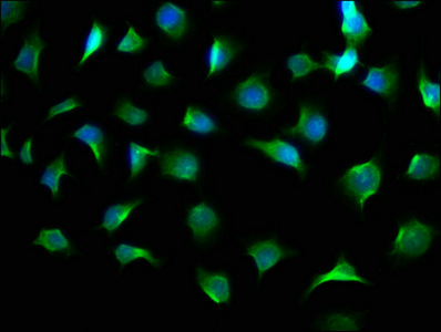
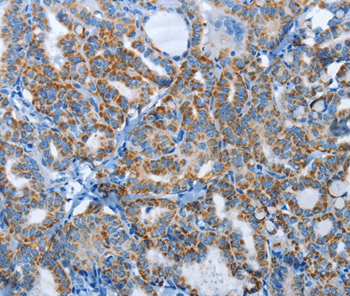
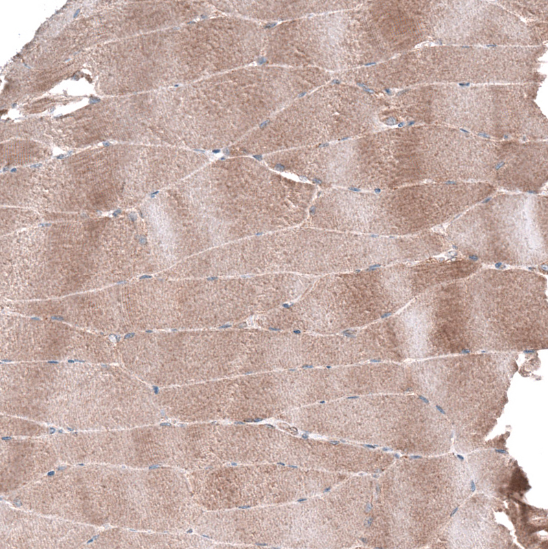
- Product NameAnti-AGL (C-term) Antibody
- Delivery Days Customer16
- ApplicationsImmunoFluorescence, Western Blot
- CertificationResearch Use Only
- ClonalityPolyclonal
- Clone IDRB4978
- Gene ID178
- Target nameAGL
- Target descriptionamylo-alpha-1,6-glucosidase and 4-alpha-glucanotransferase
- Target synonymsGDE, glycogen debranching enzyme, amylo-1, 6-glucosidase, 4-alpha-glucanotransferase, amylo-alpha-1, 6-glucosidase, 4-alpha-glucanotransferase, amylo-alpha-1, 6-glucosidase, 4-alpha-glucanotransferaseprovided, glycogen debrancher, glycogen debranching protein
- HostRabbit
- Protein IDP35573
- Protein NameGlycogen debranching enzyme
- Scientific DescriptionRabbit Anti-AGL (C-term) Antibody, 400 microl
- Storage Instruction-20°C
- UNSPSC12352203
Related products
Product group Antibodies
AGL AntibodyCSB-PA001446LA01HU
ApplicationsImmunoFluorescence, ELISA
ReactivityHuman
TargetAGL
- SizePrice
Product group Antibodies
Anti-AGL AntibodyA47232
ApplicationsImmunoHistoChemistry
ReactivityHuman
- SizePrice

Product group Antibodies
Anti-AGL Antibody Picoband(r)A02555-CARRIER-FREE
ApplicationsFlow Cytometry, ImmunoFluorescence, Western Blot, ELISA, ImmunoCytoChemistry, ImmunoHistoChemistry
ReactivityHuman, Mouse, Rat
TargetAGL
- SizePrice
Product group Antibodies
Anti-AGL AntibodyHPA028498
ApplicationsImmunoCytoChemistry, ImmunoHistoChemistry
ReactivityHuman
TargetAGL
- SizePrice
Product group Antibodies

AGL AntibodyLS-C496762
ApplicationsWestern Blot
ReactivityHuman, Mouse
TargetAGL
- SizePrice
Product group Antibodies
ApplicationsImmunoPrecipitation, Western Blot, ImmunoCytoChemistry, ImmunoHistoChemistry
ReactivityMouse, Rat
TargetAGL
- SizePrice
Product group Antibodies
ApplicationsFlow Cytometry, Western Blot, ImmunoCytoChemistry
ReactivityHuman, Mouse, Rat
TargetAGL
- SizePrice