


Figure 1. Western blot analysis of AGTR2 using anti-AGTR2 antibody (M00432). Electrophoresis was performed on a 5-20% SDS-PAGE gel at 70V (Stacking gel) / 90V (Resolving gel) for 2-3 hours. The sample well of each lane was loaded with 30 ug of sample under reducing conditions. Lane 1: human HepG2 whole cell lysates, Lane 2: human SH-SY5Y whole cell lysates, Lane 3: human Caco-2 whole cell lysates, Lane 4: rat lung tissue lysates, Lane 5: mouse lung tissue lysates. After electrophoresis, proteins were transferred to a nitrocellulose membrane at 150 mA for 50-90 minutes. Blocked the membrane with 5% non-fat milk/TBS for 1.5 hour at RT. The membrane was incubated with rabbit anti-AGTR2 antigen affinity purified monoclonal antibody (M00432) at 1:500 overnight at 4°C, then washed with TBS-0.1%Tween 3 times with 5 minutes each and probed with a goat anti-rabbit IgG-HRP secondary antibody at a dilution of 1:500 for 1.5 hour at RT. The signal is developed using an Enhanced Chemiluminescent detection (ECL) kit (Catalog # EK1002) with Tanon 5200 system. A specific band was detected for AGTR2 at approximately 45 kDa. The expected band size for AGTR2 is at 41 kDa.
Figure 1. Western blot analysis of AGTR2 using anti-AGTR2 antibody (M00432). Electrophoresis was performed on a 5-20% SDS-PAGE gel at 70V (Stacking gel) / 90V (Resolving gel) for 2-3 hours. The sample well of each lane was loaded with 30 ug of sample under reducing conditions. Lane 1: human HepG2 whole cell lysates, Lane 2: human SH-SY5Y whole cell lysates, Lane 3: human Caco-2 whole cell lysates, Lane 4: rat lung tissue lysates, Lane 5: mouse lung tissue lysates. After electrophoresis, proteins were transferred to a nitrocellulose membrane at 150 mA for 50-90 minutes. Blocked the membrane with 5% non-fat milk/TBS for 1.5 hour at RT. The membrane was incubated with rabbit anti-AGTR2 antigen affinity purified monoclonal antibody (M00432) at 1:500 overnight at 4°C, then washed with TBS-0.1%Tween 3 times with 5 minutes each and probed with a goat anti-rabbit IgG-HRP secondary antibody at a dilution of 1:500 for 1.5 hour at RT. The signal is developed using an Enhanced Chemiluminescent detection (ECL) kit (Catalog # EK1002) with Tanon 5200 system. A specific band was detected for AGTR2 at approximately 45 kDa. The expected band size for AGTR2 is at 41 kDa.
Anti-AGTR2/Angiotensin Ii Type 2 Receptor Rabbit Monoclonal Antibody
M00432
ApplicationsImmunoPrecipitation, Western Blot
Product group Antibodies
ReactivityHuman, Mouse, Rat
TargetAGTR2
Overview
- SupplierBoster Bio
- Product NameAnti-AGTR2/Angiotensin Ii Type 2 Receptor Rabbit Monoclonal Antibody
- Delivery Days Customer9
- ApplicationsImmunoPrecipitation, Western Blot
- CertificationResearch Use Only
- ClonalityMonoclonal
- Clone IDFIO-1
- Gene ID186
- Target nameAGTR2
- Target descriptionangiotensin II receptor type 2
- Target synonymsAT2, ATGR2, MRX88, type-2 angiotensin II receptor, AT2 receptor, angiotensin II type-2 receptor
- HostRabbit
- IsotypeIgG
- Protein IDP50052
- Protein NameType-2 angiotensin II receptor
- Scientific DescriptionBoster Bio Anti-AGTR2/Angiotensin Ii Type 2 Receptor Rabbit Monoclonal Antibody catalog # M00432. Tested in WB, IP applications. This antibody reacts with Human, Mouse, Rat.
- ReactivityHuman, Mouse, Rat
- Storage Instruction-20°C
- UNSPSC12352203
References
- Chang YS, Lin CL, Lee CW, et al. Exercise Normalized the Hippocampal Renin-Angiotensin System and Restored Spatial Memory Function, Neurogenesis, and Blood-Brain Barrier Permeability in the 2K1C-Hypertensive Mouse. Int J Mol Sci. 2022,23(10). doi: 10.3390/ijms23105531Read this paper
- Gomes KP, Braga PPP, de Lima CQ, et al. Antiepileptic effects of long-term intracerebroventricular infusion of angiotensin-(1-7) in an animal model of temporal lobe epilepsy. Clin Sci (Lond). 2020,134(17):2263-2277. doi: 10.1042/CS20200514Read this paper
- Macedo LM, de Ávila RI, Pedrino GR, et al. Effect of angiotensin II and angiotensin-(1-7) on proliferation of stem cells from human dental apical papilla. J Cell Physiol. 2021,236(1):366-378. doi: 10.1002/jcp.29862Read this paper
Datasheet
MSDS
Related products
Product group Antibodies
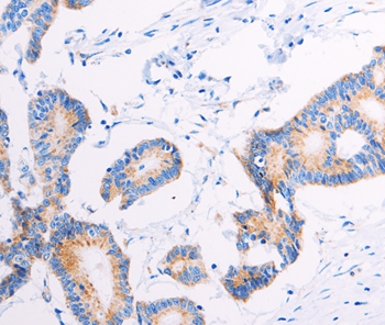
ApplicationsImmunoPrecipitation, Western Blot, ImmunoCytoChemistry, ImmunoHistoChemistry
ReactivityRat
TargetAGTR2
- SizePrice
Product group Antibodies
Anti-AGTR2 AntibodyA46124
ApplicationsImmunoHistoChemistry
ReactivityHuman
- SizePrice
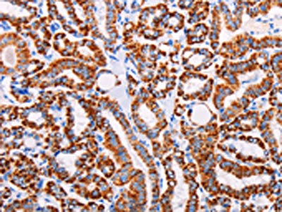
Product group Antibodies

ApplicationsImmunoHistoChemistry, ImmunoHistoChemistry Paraffin
ReactivityBovine, Canine, Equine, Hamster, Human, Mammals, Monkey, Mouse, Porcine, Rat, Sheep
TargetAGTR2
- SizePrice
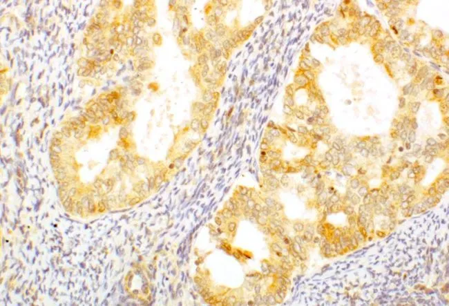
Product group Antibodies
AGTR2 AntibodyCSB-PA963716
ApplicationsELISA, ImmunoHistoChemistry
ReactivityHuman, Mouse, Rat
TargetAGTR2
- SizePrice
Product group Antibodies
AGTR2 antibodyGTX79393
ApplicationsImmunoFluorescence, Western Blot, ImmunoCytoChemistry, ImmunoHistoChemistry, ImmunoHistoChemistry Paraffin
ReactivityHuman, Rat
TargetAGTR2
- SizePrice
Product group Antibodies
AGTR2 Recombinant AntibodyBSM-61290R
ApplicationsImmunoPrecipitation, Western Blot
TargetAGTR2
- SizePrice