


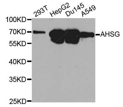
Figure 1. Western blot analysis of AHSG using anti-AHSG antibody (PB9568). Electrophoresis was performed on a 5-20% SDS-PAGE gel at 70V (Stacking gel) / 90V (Resolving gel) for 2-3 hours. The sample well of each lane was loaded with 30 ug of sample under reducing conditions. Lane 1: human placenta tissue lysates, Lane 2: human HCCT tissue lysates, Lane 3: human HCCP tissue lysates, Lane 4: rat plasma lysates. After electrophoresis, proteins were transferred to a nitrocellulose membrane at 150 mA for 50-90 minutes. Blocked the membrane with 5% non-fat milk/TBS for 1.5 hour at RT. The membrane was incubated with rabbit anti-AHSG antigen affinity purified polyclonal antibody (Catalog # PB9568) at 0.5 microg/mL overnight at 4°C, then washed with TBS-0.1%Tween 3 times with 5 minutes each and probed with a goat anti-rabbit IgG-HRP secondary antibody at a dilution of 1:5000 for 1.5 hour at RT. The signal is developed using an Enhanced Chemiluminescent detection (ECL) kit (Catalog # EK1002) with Tanon 5200 system. A specific band was detected for AHSG at approximately 55-60 kDa. The expected band size for AHSG is at 39 kDa.
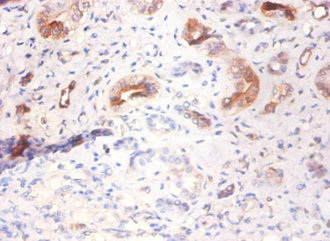
. AHSG was detected in paraffin-embedded section of Human Liver Cancer Tissue. Heat mediated antigen retrieval was performed in citrate buffer (pH6, epitope retrieval solution) for 20 mins. The tissue section was blocked with 10% goat serum. The tissue section was then incubated with 1microg/ml rabbit anti-AHSG Antibody (PB9568) overnight at 4°C. Biotinylated goat anti-rabbit IgG was used as secondary antibody and incubated for 30 minutes at 37°C. The tissue section was developed using Strepavidin-Biotin-Complex (SABC)(Catalog # SA1022) with DAB as the chromogen.")
. Overlay histogram showing HEPG2 cells stained with PB9568 (Blue line). To facilitate intracellular staining, cells were fixed with 4% paraformaldehyde and permeabilized with permeabilization buffer. The cells were blocked with 10% normal goat serum. And then incubated with rabbit anti-Fetuin A Antibody (PB9568,1microg/1x106 cells) for 30 min at 20°C. DyLight®488 conjugated goat anti-rabbit IgG (BA1127, 5-10microg/1x106 cells) was used as secondary antibody for 30 minutes at 20°C. Isotype control antibody (Green line) was rabbit IgG (1microg/1x106) used under the same conditions. Unlabelled sample (Red line) was also used as a control.")
Figure 1. Western blot analysis of AHSG using anti-AHSG antibody (PB9568). Electrophoresis was performed on a 5-20% SDS-PAGE gel at 70V (Stacking gel) / 90V (Resolving gel) for 2-3 hours. The sample well of each lane was loaded with 30 ug of sample under reducing conditions. Lane 1: human placenta tissue lysates, Lane 2: human HCCT tissue lysates, Lane 3: human HCCP tissue lysates, Lane 4: rat plasma lysates. After electrophoresis, proteins were transferred to a nitrocellulose membrane at 150 mA for 50-90 minutes. Blocked the membrane with 5% non-fat milk/TBS for 1.5 hour at RT. The membrane was incubated with rabbit anti-AHSG antigen affinity purified polyclonal antibody (Catalog # PB9568) at 0.5 microg/mL overnight at 4°C, then washed with TBS-0.1%Tween 3 times with 5 minutes each and probed with a goat anti-rabbit IgG-HRP secondary antibody at a dilution of 1:5000 for 1.5 hour at RT. The signal is developed using an Enhanced Chemiluminescent detection (ECL) kit (Catalog # EK1002) with Tanon 5200 system. A specific band was detected for AHSG at approximately 55-60 kDa. The expected band size for AHSG is at 39 kDa.
Anti-AHSG Antibody Picoband(r)
PB9568-CARRIER-FREE
ApplicationsFlow Cytometry, Western Blot, ELISA, ImmunoCytoChemistry, ImmunoHistoChemistry
Product group Antibodies
ReactivityHuman, Rat
TargetAHSG
Overview
- SupplierBoster Bio
- Product NameAnti-AHSG Antibody Picoband(r)
- Delivery Days Customer9
- Application Supplier NoteTested Species: In-house tested species with positive results. By Heat: Boiling the paraffin sections in 10mM citrate buffer, pH6.0, for 20mins is required for the staining of formalin/paraffin sections. Other applications have not been tested. Optimal dilutions should be determined by end users.
- ApplicationsFlow Cytometry, Western Blot, ELISA, ImmunoCytoChemistry, ImmunoHistoChemistry
- CertificationResearch Use Only
- ClonalityPolyclonal
- Concentration500 ug/ml
- Gene ID197
- Target nameAHSG
- Target descriptionalpha 2-HS glycoprotein
- Target synonymsA2HS, AHS, APMR1, FETUA, HSGA, alpha-2-HS-glycoprotein, alpha-2-Z-globulin, ba-alpha-2-glycoprotein, fetuin-A
- HostRabbit
- IsotypeIgG
- Protein IDP02765
- Protein NameAlpha-2-HS-glycoprotein
- Scientific DescriptionBoster Bio Anti-AHSG Antibody Picoband® catalog # PB9568. Tested in ELISA, Flow Cytometry, IHC, ICC, WB applications. This antibody reacts with Human, Rat. The brand Picoband indicates this is a premium antibody that guarantees superior quality, high affinity, and strong signals with minimal background in Western blot applications. Only our best-performing antibodies are designated as Picoband, ensuring unmatched performance.
- ReactivityHuman, Rat
- Storage Instruction-20°C,2°C to 8°C
- UNSPSC12352203
Related products
Product group Antibodies
AHSG AntibodyCSB-PA06369A0RB
ApplicationsImmunoFluorescence, ELISA, ImmunoHistoChemistry
ReactivityHuman
TargetAHSG
- SizePrice
Product group Antibodies
Anti-AHSG Antibody144-01647
ApplicationsImmunoFluorescence, Western Blot
ReactivityHuman
TargetAHSG
- SizePrice
Product group Antibodies
Anti-AHSG AntibodyA29771
ApplicationsImmunoFluorescence, Western Blot, ImmunoHistoChemistry
ReactivityHuman
- SizePrice

Product group Antibodies
Anti-AHSG AntibodyHPA001524
ApplicationsWestern Blot, ImmunoCytoChemistry, ImmunoHistoChemistry
ReactivityHuman
TargetAHSG
- SizePrice
Product group Antibodies

Goat anti-AHSGEB12227
ApplicationsWestern Blot, ELISA
ReactivityHuman
TargetAHSG
- SizePrice
Product group Antibodies

AHSG / Fetuin A AntibodyLS-C401227
ApplicationsWestern Blot, ELISA, ImmunoHistoChemistry
ReactivityHuman
TargetAHSG
- SizePrice
Product group Antibodies
ApplicationsWestern Blot, ELISA, ImmunoHistoChemistry, ImmunoHistoChemistry Paraffin
ReactivityBovine, Canine, Equine, Human, Mouse, Rabbit, Rat, Sheep
TargetAHSG
- SizePrice

Product group Antibodies
Fetuin A antibodyGTX30034
ApplicationsImmunoFluorescence, Western Blot, ImmunoCytoChemistry
ReactivityHuman
TargetAHSG
- SizePrice