


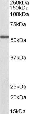
Figure 1. Western blot analysis of ALDH6A1 using anti-ALDH6A1 antibody (A08868-1). Electrophoresis was performed on a 5-20% SDS-PAGE gel at 70V (Stacking gel) / 90V (Resolving gel) for 2-3 hours. The sample well of each lane was loaded with 50ug of sample under reducing conditions. Lane 1: rat liver tissue lysates, Lane 2: rat heart tissue lysates, Lane 3: rat kidney tissue lysates, Lane 4: mouse liver tissue lysates, Lane 5: mouse kidney tissue lysates, Lane 6: mouse NIH/3T3 whole cell lysates, Lane 7: human MCF-7 whole cell lysates, Lane 8: human HEPG2 whole cell lysates. After Electrophoresis, proteins were transferred to a Nitrocellulose membrane at 150mA for 50-90 minutes. Blocked the membrane with 5% Non-fat Milk/ TBS for 1.5 hour at RT. The membrane was incubated with rabbit anti-ALDH6A1 antigen affinity purified polyclonal antibody (Catalog # A08868-1) at 0.25 microg/mL overnight at 4°C, then washed with TBS-0.1%Tween 3 times with 5 minutes each and probed with a goat anti-rabbit IgG-HRP secondary antibody at a dilution of 1:5000 for 1.5 hour at RT. The signal is developed using an Enhanced Chemiluminescent detection (ECL) kit (Catalog # EK1002) with Tanon 5200 system. A specific band was detected for ALDH6A1 at approximately 58KD. The expected band size for ALDH6A1 is at 58KD.
. ALDH6A1 was detected in paraffin-embedded section of human liver cancer tissue. Heat mediated antigen retrieval was performed in EDTA buffer (pH8.0, epitope retrieval solution). The tissue section was blocked with 10% goat serum. The tissue section was then incubated with 2microg/ml rabbit anti-ALDH6A1 Antibody (A08868-1) overnight at 4°C. Biotinylated goat anti-rabbit IgG was used as secondary antibody and incubated for 30 minutes at 37°C. The tissue section was developed using Strepavidin-Biotin-Complex (SABC) (Catalog # SA1022) with DAB as the chromogen.")
Figure 1. Western blot analysis of ALDH6A1 using anti-ALDH6A1 antibody (A08868-1). Electrophoresis was performed on a 5-20% SDS-PAGE gel at 70V (Stacking gel) / 90V (Resolving gel) for 2-3 hours. The sample well of each lane was loaded with 50ug of sample under reducing conditions. Lane 1: rat liver tissue lysates, Lane 2: rat heart tissue lysates, Lane 3: rat kidney tissue lysates, Lane 4: mouse liver tissue lysates, Lane 5: mouse kidney tissue lysates, Lane 6: mouse NIH/3T3 whole cell lysates, Lane 7: human MCF-7 whole cell lysates, Lane 8: human HEPG2 whole cell lysates. After Electrophoresis, proteins were transferred to a Nitrocellulose membrane at 150mA for 50-90 minutes. Blocked the membrane with 5% Non-fat Milk/ TBS for 1.5 hour at RT. The membrane was incubated with rabbit anti-ALDH6A1 antigen affinity purified polyclonal antibody (Catalog # A08868-1) at 0.25 microg/mL overnight at 4°C, then washed with TBS-0.1%Tween 3 times with 5 minutes each and probed with a goat anti-rabbit IgG-HRP secondary antibody at a dilution of 1:5000 for 1.5 hour at RT. The signal is developed using an Enhanced Chemiluminescent detection (ECL) kit (Catalog # EK1002) with Tanon 5200 system. A specific band was detected for ALDH6A1 at approximately 58KD. The expected band size for ALDH6A1 is at 58KD.
Anti-ALDH6A1 Antibody Picoband(r)
A08868-1-CARRIER-FREE
ApplicationsWestern Blot, ImmunoHistoChemistry
Product group Antibodies
ReactivityHuman, Mouse, Rat
TargetALDH6A1
Overview
- SupplierBoster Bio
- Product NameAnti-ALDH6A1 Antibody Picoband(r)
- Delivery Days Customer9
- ApplicationsWestern Blot, ImmunoHistoChemistry
- CertificationResearch Use Only
- ClonalityPolyclonal
- Concentration500 ug/ml
- Gene ID4329
- Target nameALDH6A1
- Target descriptionaldehyde dehydrogenase 6 family member A1
- Target synonymsMMSADHA, MMSDH, methylmalonate-semialdehyde/malonate-semialdehyde dehydrogenase [acylating], mitochondrial, malonate-semialdehyde dehydrogenase (acetylating), methylmalonate-semialdehyde dehydrogenase [acylating], mitochondrial, mitochondrial acylating methylmalonate-semialdehyde dehydrogenase, testicular tissue protein Li 122
- HostRabbit
- IsotypeIgG
- Protein IDQ02252
- Protein NameMethylmalonate-semialdehyde/malonate-semialdehyde dehydrogenase [acylating], mitochondrial
- Scientific DescriptionBoster Bio Anti-ALDH6A1 Antibody Picoband® catalog # A08868-1. Tested in IHC, WB applications. This antibody reacts with Human, Mouse, Rat. The brand Picoband indicates this is a premium antibody that guarantees superior quality, high affinity, and strong signals with minimal background in Western blot applications. Only our best-performing antibodies are designated as Picoband, ensuring unmatched performance.
- ReactivityHuman, Mouse, Rat
- Storage Instruction-20°C,2°C to 8°C
- UNSPSC12352203
Related products
Product group Antibodies
ApplicationsWestern Blot, ELISA
ReactivityHuman, Mouse, Rat
- SizePrice
Product group Antibodies
Anti-ALDH6A1 Antibody144-03309
ApplicationsWestern Blot
ReactivityHuman, Mouse, Rat
TargetALDH6A1
- SizePrice

Product group Antibodies
ALDH6A1 (7C4) Monoclonal AntibodyBSM-51430M
ApplicationsImmunoFluorescence, Western Blot, ImmunoHistoChemistry, ImmunoHistoChemistry Paraffin
ReactivityHuman
TargetALDH6A1
- SizePrice
Product group Antibodies
ALDH6A1 AntibodyCSB-PA001578LA01HU
ApplicationsWestern Blot, ELISA, ImmunoHistoChemistry
ReactivityHuman, Mouse
TargetALDH6A1
- SizePrice
Product group Antibodies

ApplicationsWestern Blot, ELISA, ImmunoHistoChemistry
ReactivityBovine, Canine, Human, Mouse, Porcine, Rat
TargetALDH6A1
- SizePrice
Product group Antibodies
ALDH6A1 Polyclonal AntibodyCAC14675
ApplicationsWestern Blot, ELISA, ImmunoHistoChemistry
ReactivityMouse
TargetALDH6A1
- SizePrice
Product group Antibodies

ALDH6A1 AntibodyLS-C401217
ApplicationsWestern Blot, ELISA, ImmunoHistoChemistry
ReactivityHuman, Mouse, Rat
TargetALDH6A1
- SizePrice
Product group Antibodies
ALDH6A1 antibodyGTX103405
ApplicationsWestern Blot, ImmunoHistoChemistry, ImmunoHistoChemistry Paraffin
ReactivityHuman, Mouse
TargetALDH6A1
- SizePrice

Product group Antibodies
Anti-ALDH6A1 AntibodyHPA029072
ApplicationsWestern Blot, ImmunoHistoChemistry
ReactivityHuman, Mouse, Rat
TargetALDH6A1
- SizePrice