


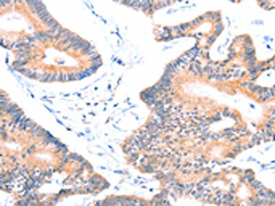
Immunohistochemical staining of human kidney shows moderate to strong membranous positivity in cells in tubules.
![Lane 1: Marker [kDa] 230, 130, 95, 72, 56, 36, 28, 17, 11. Lane 2: Human cell line RT-4. Lane 3: Human cell line U-251MG sp](https://atlasantibodies.s3.amazonaws.com/images/wb/hpa031033-wb-1.jpg "Lane 1: Marker [kDa] 230, 130, 95, 72, 56, 36, 28, 17, 11. Lane 2: Human cell line RT-4. Lane 3: Human cell line U-251MG sp")
Immunohistochemical staining of human kidney shows moderate to strong membranous positivity in cells in tubules.
Anti-AOC1 Antibody
HPA031033
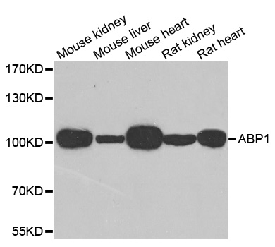
ApplicationsWestern Blot, ImmunoHistoChemistry
Product group Antibodies
ReactivityHuman
TargetAOC1
Overview
- SupplierAtlas Antibodies
- Product NameAnti-AOC1 Antibody
- Delivery Days Customer4
- ApplicationsWestern Blot, ImmunoHistoChemistry
- CertificationResearch Use Only
- ClonalityPolyclonal
- ConjugateUnconjugated
- Gene ID26
- Target nameAOC1
- Target descriptionamine oxidase copper containing 1
- Target synonymsABP, ABP1, DAO, DAO1, KAO, KDAO, diamine oxidase [copper-containing], amiloride binding protein 1 (amine oxidase (copper-containing)), amiloride-binding protein 1, amiloride-sensitive amine oxidase, amine oxidase copper domain-containing protein 1, diamine oxidase, histaminase, kidney amine oxidase
- HostRabbit
- IsotypeIgG
- Protein IDP19801
- Protein NameDiamine oxidase [copper-containing]
- Scientific DescriptionRecombinant Protein Epitope Signature Tag (PrEST) antigen sequence
- ReactivityHuman
- Storage Instruction-20°C,2°C to 8°C
- UNSPSC41116161
Datasheet
MSDS
Related products

Product group Antibodies
Anti-ABP1/AOC1 Antibody Picoband(r)A08386-1-CARRIER-FREE
ApplicationsFlow Cytometry, ImmunoFluorescence, Western Blot, ELISA, ImmunoCytoChemistry, ImmunoHistoChemistry
ReactivityHuman, Monkey
TargetAOC1
- SizePrice
Product group Antibodies
Anti-AOC1 Antibody144-06249
ApplicationsImmunoFluorescence, Western Blot
ReactivityHuman, Mouse, Rat
TargetAOC1
- SizePrice
Product group Antibodies
Anti-KAO AntibodyA31177
ApplicationsWestern Blot, ImmunoHistoChemistry
ReactivityHuman, Mouse, Rat
- SizePrice

Product group Antibodies
Anti-AOC1 AntibodyHPA031032
ApplicationsWestern Blot, ImmunoHistoChemistry
ReactivityHuman
TargetAOC1
- SizePrice
Product group Antibodies
Anti-AOC1 AntibodyHPA031032
ApplicationsWestern Blot, ImmunoHistoChemistry
ReactivityHuman
TargetAOC1
- SizePrice
Product group Antibodies
AOC1 AntibodyCSB-PA104599
ApplicationsELISA, ImmunoHistoChemistry
ReactivityHuman, Mouse, Rat
TargetAOC1
- SizePrice
Product group Antibodies

AOC1 AntibodyLS-C482545
ApplicationsWestern Blot
ReactivityHuman, Mouse, Rat
TargetAOC1
- SizePrice