

Anti-AP2B1 Antibody
A283440
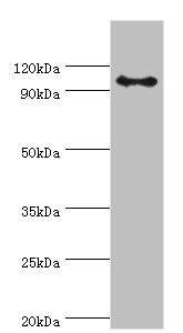
ApplicationsWestern Blot
Product group Antibodies
ReactivityHuman, Mouse, Rat
Overview
- SupplierAntibodies.com
- Product NameAnti-AP2B1 Antibody
- Delivery Days Customer7
- ApplicationsWestern Blot
- CertificationResearch Use Only
- ClonalityPolyclonal
- ConjugateUnconjugated
- HostRabbit
- IsotypeIgG
- Scientific DescriptionRabbit polyclonal antibody to AP2B1.
- ReactivityHuman, Mouse, Rat
- Storage Instruction-20°C
- UNSPSC12352203
Related products
Product group Antibodies
Anti-AP2B1 Antibody144-01995
ApplicationsWestern Blot, ImmunoHistoChemistry
ReactivityHuman, Mouse
TargetAP2B1
- SizePrice
Product group Antibodies
AP2B1 antibodyGTX112479
ApplicationsWestern Blot, ImmunoHistoChemistry, ImmunoHistoChemistry Paraffin
ReactivityHuman, Mouse
TargetAP2B1
- SizePrice
Product group Antibodies

AP2B1 AntibodyLS-C331812
ApplicationsWestern Blot, ImmunoHistoChemistry
ReactivityHuman, Mouse
TargetAP2B1
- SizePrice

Product group Antibodies
Anti-AP2B1 AntibodyHPA056733
ApplicationsWestern Blot, ImmunoCytoChemistry
ReactivityHuman
TargetAP2B1
- SizePrice
Product group Antibodies
AP2B1 AntibodyCSB-PA001871ESR1HU
ApplicationsWestern Blot, ELISA, ImmunoHistoChemistry
ReactivityHuman, Mouse
TargetAP2B1
- SizePrice
Product group Antibodies
Anti-AP2B1Y158120
ApplicationsWestern Blot, ELISA, ImmunoHistoChemistry
ReactivityHuman, Mouse, Rat
- SizePrice

Product group Antibodies
Anti-AP2B1 Antibody Picoband(r)A06544-CARRIER-FREE
ApplicationsWestern Blot
ReactivityHuman, Mouse, Rat
TargetAP2B1
- SizePrice