


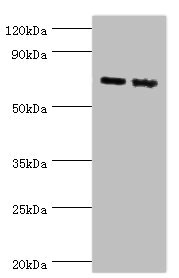
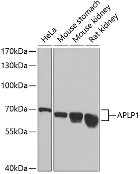
Figure 1. Western blot analysis of APLP1 using anti-APLP1 antibody (A03929). Electrophoresis was performed on a 5-20% SDS-PAGE gel at 70V (Stacking gel) / 90V (Resolving gel) for 2-3 hours. The sample well of each lane was loaded with 30 ug of sample under reducing conditions. Lane 1: human SH-SY5Y whole cell lysates, Lane 2: human U-87MG whole cell lysates, Lane 3: rat brain tissue lysates, Lane 4: rat liver tissue lysates, Lane 5: rat RH35 whole cell lysates, Lane 6: mouse brain tissue lysates, Lane 7: mouse Neuro-2a whole cell lysates. After electrophoresis, proteins were transferred to a nitrocellulose membrane at 150 mA for 50-90 minutes. Blocked the membrane with 5% non-fat milk/TBS for 1.5 hour at RT. The membrane was incubated with rabbit anti-APLP1 antigen affinity purified polyclonal antibody (Catalog # A03929) at 0.5 microg/mL overnight at 4°C, then washed with TBS-0.1%Tween 3 times with 5 minutes each and probed with a goat anti-rabbit IgG-HRP secondary antibody at a dilution of 1:5000 for 1.5 hour at RT. The signal is developed using an Enhanced Chemiluminescent detection (ECL) kit (Catalog # EK1002) with Tanon 5200 system. A specific band was detected for APLP1 at approximately 72 kDa. The expected band size for APLP1 is at 72,97 kDa.
. Overlay histogram showing SiHa cells stained with A03929 (Blue line). To facilitate intracellular staining, cells were fixed with 4% paraformaldehyde and permeabilized with permeabilization buffer. The cells were blocked with 10% normal goat serum. And then incubated with rabbit anti-APLP1 Antibody (A03929, 1 microg/1x106 cells) for 30 min at 20°C. DyLight®488 conjugated goat anti-rabbit IgG (BA1127, 5-10 microg/1x106 cells) was used as secondary antibody for 30 minutes at 20°C. Isotype control antibody (Green line) was rabbit IgG (1 microg/1x106) used under the same conditions. Unlabelled sample (Red line) was also used as a control.")
Figure 1. Western blot analysis of APLP1 using anti-APLP1 antibody (A03929). Electrophoresis was performed on a 5-20% SDS-PAGE gel at 70V (Stacking gel) / 90V (Resolving gel) for 2-3 hours. The sample well of each lane was loaded with 30 ug of sample under reducing conditions. Lane 1: human SH-SY5Y whole cell lysates, Lane 2: human U-87MG whole cell lysates, Lane 3: rat brain tissue lysates, Lane 4: rat liver tissue lysates, Lane 5: rat RH35 whole cell lysates, Lane 6: mouse brain tissue lysates, Lane 7: mouse Neuro-2a whole cell lysates. After electrophoresis, proteins were transferred to a nitrocellulose membrane at 150 mA for 50-90 minutes. Blocked the membrane with 5% non-fat milk/TBS for 1.5 hour at RT. The membrane was incubated with rabbit anti-APLP1 antigen affinity purified polyclonal antibody (Catalog # A03929) at 0.5 microg/mL overnight at 4°C, then washed with TBS-0.1%Tween 3 times with 5 minutes each and probed with a goat anti-rabbit IgG-HRP secondary antibody at a dilution of 1:5000 for 1.5 hour at RT. The signal is developed using an Enhanced Chemiluminescent detection (ECL) kit (Catalog # EK1002) with Tanon 5200 system. A specific band was detected for APLP1 at approximately 72 kDa. The expected band size for APLP1 is at 72,97 kDa.
Anti-APLP1 Antibody Picoband(r)
A03929-CARRIER-FREE
ApplicationsFlow Cytometry, Western Blot, ELISA
Product group Antibodies
ReactivityHuman, Mouse, Rat
TargetAPLP1
Overview
- SupplierBoster Bio
- Product NameAnti-APLP1 Antibody Picoband(r)
- Delivery Days Customer9
- Application Supplier NoteTested Species: In-house tested species with positive results. Other applications have not been tested. Optimal dilutions should be determined by end users.
- ApplicationsFlow Cytometry, Western Blot, ELISA
- CertificationResearch Use Only
- ClonalityPolyclonal
- Concentration500 ug/ml
- Gene ID333
- Target nameAPLP1
- Target descriptionamyloid beta precursor like protein 1
- Target synonymsAPLP, amyloid beta precursor like protein 1, amyloid beta (A4) precursor-like protein 1, amyloid precursor-like protein 1, amyloid-like protein 1
- HostRabbit
- IsotypeIgG
- Protein IDP51693
- Protein NameAmyloid beta precursor like protein 1
- Scientific DescriptionBoster Bio Anti-APLP1 Antibody Picoband® catalog # A03929. Tested in ELISA, Flow Cytometry, WB applications. This antibody reacts with Human, Mouse, Rat. The brand Picoband indicates this is a premium antibody that guarantees superior quality, high affinity, and strong signals with minimal background in Western blot applications. Only our best-performing antibodies are designated as Picoband, ensuring unmatched performance.
- ReactivityHuman, Mouse, Rat
- Storage Instruction-20°C,2°C to 8°C
- UNSPSC12352203
Related products
Product group Antibodies
APLP1 AntibodyCSB-PA001911ESR1HU
ApplicationsWestern Blot, ELISA
ReactivityHuman, Mouse
TargetAPLP1
- SizePrice
Product group Antibodies
Aplp1 Polyclonal AntibodyCAC10592
ApplicationsWestern Blot, ELISA
ReactivityMouse
TargetAPLP1
- SizePrice
Product group Antibodies
Anti-APLP1 Antibody144-06870
ApplicationsImmunoFluorescence, Western Blot
ReactivityHuman, Mouse, Rat
TargetAPLP1
- SizePrice
Product group Antibodies
Anti-APLP1 AntibodyA30596
ApplicationsWestern Blot, ImmunoHistoChemistry
ReactivityHuman, Mouse, Rat
- SizePrice
Product group Antibodies
Anti-APLP1 AntibodyHPA028970
ApplicationsWestern Blot, ImmunoHistoChemistry
ReactivityHuman
TargetAPLP1
- SizePrice
Product group Antibodies

APLP1 / APLP-1 AntibodyLS-C400463
ApplicationsWestern Blot, ELISA, ImmunoHistoChemistry
ReactivityHuman, Mouse
TargetAPLP1
- SizePrice
Product group Antibodies
APLP1 Polyclonal AntibodyBS-8465R
ApplicationsImmunoFluorescence, Western Blot, ELISA, ImmunoCytoChemistry, ImmunoHistoChemistry, ImmunoHistoChemistry Frozen, ImmunoHistoChemistry Paraffin
ReactivityBovine, Canine, Equine, Human, Mouse, Rat, Sheep
TargetAPLP1
- SizePrice
Product group Antibodies
APLP1 antibodyGTX30055
ApplicationsWestern Blot, ImmunoHistoChemistry, ImmunoHistoChemistry Paraffin
ReactivityHuman, Mouse, Rat
TargetAPLP1
- SizePrice