


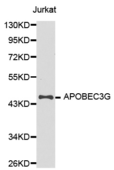
Figure 1. Western blot analysis of APOBEC3G using anti-APOBEC3G antibody (PB9985). Electrophoresis was performed on a 5-20% SDS-PAGE gel at 70V (Stacking gel) / 90V (Resolving gel) for 2-3 hours. The sample well of each lane was loaded with 30 ug of sample under reducing conditions. Lane 1: human Jurkat whole cell lysates, Lane 2: human HEL whole cell lysates, Lane 3: human RT4 whole cell lysates, Lane 4: rat testis tissue lysates, Lane 5: rat PC-12 whole cell lysates, Lane 6: mouse testis tissue lysates, Lane 7: mouse RAW264.7 whole cell lysates. After electrophoresis, proteins were transferred to a nitrocellulose membrane at 150 mA for 50-90 minutes. Blocked the membrane with 5% non-fat milk/TBS for 1.5 hour at RT. The membrane was incubated with rabbit anti-APOBEC3G antigen affinity purified polyclonal antibody (Catalog # PB9985) at 0.5 microg/mL overnight at 4°C, then washed with TBS-0.1%Tween 3 times with 5 minutes each and probed with a goat anti-rabbit IgG-HRP secondary antibody at a dilution of 1:5000 for 1.5 hour at RT. The signal is developed using an Enhanced Chemiluminescent detection (ECL) kit (Catalog # EK1002) with Tanon 5200 system. A specific band was detected for APOBEC3G at approximately 46 kDa. The expected band size for APOBEC3G is at 46 kDa.
. APOBEC3G was detected in an immunocytochemical section of A431 cells. Enzyme antigen retrieval was performed using IHC enzyme antigen retrieval reagent (AR0022) for 15 mins. The cells were blocked with 10% goat serum. And then incubated with 5 microg/mL rabbit anti-APOBEC3G Antibody (PB9985) overnight at 4°C. DyLight®488 Conjugated Goat Anti-Rabbit IgG (BA1127) was used as secondary antibody at 1:500 dilution and incubated for 30 minutes at 37°C. The section was counterstained with DAPI. Visualize using a fluorescence microscope and filter sets appropriate for the label used.")
Figure 1. Western blot analysis of APOBEC3G using anti-APOBEC3G antibody (PB9985). Electrophoresis was performed on a 5-20% SDS-PAGE gel at 70V (Stacking gel) / 90V (Resolving gel) for 2-3 hours. The sample well of each lane was loaded with 30 ug of sample under reducing conditions. Lane 1: human Jurkat whole cell lysates, Lane 2: human HEL whole cell lysates, Lane 3: human RT4 whole cell lysates, Lane 4: rat testis tissue lysates, Lane 5: rat PC-12 whole cell lysates, Lane 6: mouse testis tissue lysates, Lane 7: mouse RAW264.7 whole cell lysates. After electrophoresis, proteins were transferred to a nitrocellulose membrane at 150 mA for 50-90 minutes. Blocked the membrane with 5% non-fat milk/TBS for 1.5 hour at RT. The membrane was incubated with rabbit anti-APOBEC3G antigen affinity purified polyclonal antibody (Catalog # PB9985) at 0.5 microg/mL overnight at 4°C, then washed with TBS-0.1%Tween 3 times with 5 minutes each and probed with a goat anti-rabbit IgG-HRP secondary antibody at a dilution of 1:5000 for 1.5 hour at RT. The signal is developed using an Enhanced Chemiluminescent detection (ECL) kit (Catalog # EK1002) with Tanon 5200 system. A specific band was detected for APOBEC3G at approximately 46 kDa. The expected band size for APOBEC3G is at 46 kDa.
Anti-APOBEC3G Antibody Picoband(r)
PB9985-CARRIER-FREE
ApplicationsImmunoFluorescence, Western Blot, ImmunoCytoChemistry
Product group Antibodies
ReactivityHuman, Mouse, Rat
TargetAPOBEC3G
Overview
- SupplierBoster Bio
- Product NameAnti-APOBEC3G Antibody Picoband(r)
- Delivery Days Customer9
- Application Supplier NoteTested Species: In-house tested species with positive results. By Heat: Boiling the paraffin sections in 10mM citrate buffer, pH6.0, for 20mins is required for the staining of formalin/paraffin sections. Other applications have not been tested. Optimal dilutions should be determined by end users.
- ApplicationsImmunoFluorescence, Western Blot, ImmunoCytoChemistry
- CertificationResearch Use Only
- ClonalityPolyclonal
- Concentration500 ug/ml
- Gene ID60489
- Target nameAPOBEC3G
- Target descriptionapolipoprotein B mRNA editing enzyme catalytic subunit 3G
- Target synonymsA3G, ARCD, ARP-9, ARP9, CEM-15, CEM15, MDS019, bK150C2.7, dJ494G10.1, DNA dC->dU-editing enzyme APOBEC-3G, APOBEC-related cytidine deaminase, APOBEC-related protein 9, DNA dC->dU editing enzyme, apolipoprotein B editing enzyme catalytic polypeptide-like 3G, apolipoprotein B mRNA editing enzyme cytidine deaminase, apolipoprotein B mRNA editing enzyme, catalytic polypeptide-like 3G, apolipoprotein B mRNA-editing enzyme catalytic polypeptide 3G, deoxycytidine deaminase, phorbolin-like protein MDS019
- HostRabbit
- IsotypeIgG
- Protein IDQ9HC16
- Protein NameDNA dC->dU-editing enzyme APOBEC-3G
- Scientific DescriptionBoster Bio Anti-APOBEC3G Antibody Picoband® catalog # PB9985. Tested in IF, ICC, WB applications. This antibody reacts with Human, Mouse, Rat. The brand Picoband indicates this is a premium antibody that guarantees superior quality, high affinity, and strong signals with minimal background in Western blot applications. Only our best-performing antibodies are designated as Picoband, ensuring unmatched performance.
- ReactivityHuman, Mouse, Rat
- Storage Instruction-20°C,2°C to 8°C
- UNSPSC12352203
Related products
Product group Antibodies
Anti-APOBEC3G Antibody144-62833
ApplicationsImmunoFluorescence, Western Blot, ImmunoHistoChemistry
ReactivityHuman, Rat
TargetAPOBEC3G
- SizePrice
Product group Antibodies
Anti-APOBEC3G AntibodyA34811
ApplicationsImmunoFluorescence, Western Blot, ImmunoHistoChemistry
ReactivityHuman
- SizePrice
Product group Antibodies
APOBEC3G Polyclonal AntibodyBS-15407R
ApplicationsImmunoFluorescence, Western Blot, ELISA, ImmunoCytoChemistry, ImmunoHistoChemistry, ImmunoHistoChemistry Frozen, ImmunoHistoChemistry Paraffin
ReactivityHuman
TargetAPOBEC3G
- SizePrice
Product group Antibodies
APOBEC3G AntibodyCSB-PA001926LA01HU
ApplicationsImmunoFluorescence, ELISA, ImmunoHistoChemistry
ReactivityHuman
TargetAPOBEC3G
- SizePrice
Product group Antibodies

Goat anti-APOBEC3G / ARP9EB06714
ApplicationsELISA, ImmunoHistoChemistry
ReactivityHuman
TargetAPOBEC3G
- SizePrice
Product group Antibodies

APOBEC3G / CEM15 AntibodyLS-C401263
ApplicationsELISA, ImmunoHistoChemistry
ReactivityHuman
TargetAPOBEC3G
- SizePrice

Product group Antibodies
Anti-APOBEC3G AntibodyHPA001812
ApplicationsImmunoHistoChemistry
ReactivityHuman
TargetAPOBEC3G
- SizePrice
Product group Antibodies
APOBEC3G antibody [N1C1]GTX112176
ApplicationsWestern Blot
ReactivityHuman
TargetAPOBEC3G
- SizePrice