


Figure 2. IHC analysis of Apolipoprotein CIII using anti-Apolipoprotein CIII antibody (PB10042). Apolipoprotein CIII was detected in a paraffin-embedded section of human liver cancer tissue. Heat mediated antigen retrieval was performed in EDTA buffer (pH 8.0, epitope retrieval solution). The tissue section was blocked with 10% goat serum. The tissue section was then incubated with 1 microg/ml rabbit anti-Apolipoprotein CIII Antibody (PB10042) overnight at 4°C. Biotinylated goat anti-rabbit IgG was used as secondary antibody and incubated for 30 minutes at 37°C. The tissue section was developed using Strepavidin-Biotin-Complex (SABC) (Catalog # SA1022) with DAB as the chromogen.
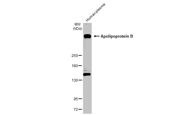
. Electrophoresis was performed on a 5-20% SDS-PAGE gel at 70V (Stacking gel) / 90V (Resolving gel) for 2-3 hours. The sample well of each lane was loaded with 30 ug of sample under reducing conditions. Lane 1: HELA whole cell lysates. After electrophoresis, proteins were transferred to a nitrocellulose membrane at 150 mA for 50-90 minutes. Blocked the membrane with 5% non-fat milk/TBS for 1.5 hour at RT. The membrane was incubated with rabbit anti-Apolipoprotein CIII antigen affinity purified polyclonal antibody (Catalog # PB10042) at 0.5 microg/mL overnight at 4°C, then washed with TBS-0.1%Tween 3 times with 5 minutes each and probed with a goat anti-rabbit IgG-HRP secondary antibody at a dilution of 1:5000 for 1.5 hour at RT. The signal is developed using an Enhanced Chemiluminescent detection (ECL) kit (Catalog # EK1002) with Tanon 5200 system. A specific band was detected for Apolipoprotein CIII at approximately 250 kDa. The expected band size for Apolipoprotein CIII is at 516 kDa.")
Figure 2. IHC analysis of Apolipoprotein CIII using anti-Apolipoprotein CIII antibody (PB10042). Apolipoprotein CIII was detected in a paraffin-embedded section of human liver cancer tissue. Heat mediated antigen retrieval was performed in EDTA buffer (pH 8.0, epitope retrieval solution). The tissue section was blocked with 10% goat serum. The tissue section was then incubated with 1 microg/ml rabbit anti-Apolipoprotein CIII Antibody (PB10042) overnight at 4°C. Biotinylated goat anti-rabbit IgG was used as secondary antibody and incubated for 30 minutes at 37°C. The tissue section was developed using Strepavidin-Biotin-Complex (SABC) (Catalog # SA1022) with DAB as the chromogen.
Anti-Apolipoprotein B/APOB Antibody Picoband(r)
PB10042-CARRIER-FREE
ApplicationsWestern Blot, ImmunoHistoChemistry
Product group Antibodies
ReactivityHuman
TargetAPOB
Overview
- SupplierBoster Bio
- Product NameAnti-Apolipoprotein B/APOB Antibody Picoband(r)
- Delivery Days Customer9
- Application Supplier NoteTested Species: In-house tested species with positive results. By Heat: Boiling the paraffin sections in 10mM citrate buffer, pH6.0, for 20mins is required for the staining of formalin/paraffin sections. Other applications have not been tested. Optimal dilutions should be determined by end users.
- ApplicationsWestern Blot, ImmunoHistoChemistry
- CertificationResearch Use Only
- ClonalityPolyclonal
- Concentration500 ug/ml
- Gene ID338
- Target nameAPOB
- Target descriptionapolipoprotein B
- Target synonymsFCHL2, FLDB, LDLCQ4, apoB-100, apoB-48, apolipoprotein B-100, apolipoprotein B (including Ag(x) antigen), apolipoprotein B48
- HostRabbit
- IsotypeIgG
- Protein IDP04114
- Protein NameApolipoprotein B-100
- Scientific DescriptionBoster Bio Anti-Apolipoprotein B/APOB Antibody Picoband® catalog # PB10042. Tested in IHC, WB applications. This antibody reacts with Human. The brand Picoband indicates this is a premium antibody that guarantees superior quality, high affinity, and strong signals with minimal background in Western blot applications. Only our best-performing antibodies are designated as Picoband, ensuring unmatched performance.
- ReactivityHuman
- Storage Instruction-20°C,2°C to 8°C
- UNSPSC12352203
Related products

Product group Antibodies
ApplicationsWestern Blot, ELISA
ReactivityHuman, Primate
TargetAPOB
- SizePrice
Product group Antibodies
Anti-APOB Antibody144-01330
ApplicationsWestern Blot
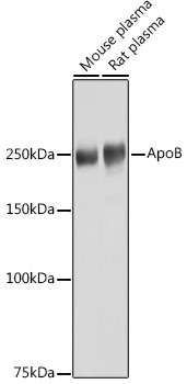
ReactivityHuman, Mouse, Rat
TargetAPOB
- SizePrice
Product group Antibodies

Anti-Apolipoprotein B-100 [MabB9]AB03939-10.0
ApplicationsWestern Blot, ELISA
ReactivityHuman
TargetAPOB
- SizePrice
Product group Antibodies
ApplicationsImmunoFluorescence, Western Blot, ImmunoCytoChemistry
ReactivityHuman, Mouse, Rat
- SizePrice
Product group Antibodies
ApoB Polyclonal AntibodyBS-6333R
ApplicationsWestern Blot
ReactivityCanine, Equine, Human, Mouse, Rabbit, Rat
TargetAPOB
- SizePrice
Product group Antibodies
APOB AntibodyCSB-PA001918LA01HU
ApplicationsELISA
ReactivityHuman
TargetAPOB
- SizePrice
Product group Antibodies

Goat anti-APOBEB08792
ApplicationsWestern Blot, ELISA, ImmunoHistoChemistry
ReactivityHuman
TargetAPOB
- SizePrice
Product group Antibodies
ApplicationsImmunoPrecipitation, Western Blot, ImmunoCytoChemistry, ImmunoHistoChemistry
TargetAPOB
- SizePrice
Product group Antibodies
Apolipoprotein B antibodyGTX135994
ApplicationsWestern Blot, ELISA, ImmunoHistoChemistry, ImmunoHistoChemistry Paraffin
ReactivityHuman, Mouse
TargetAPOB
- SizePrice
Product group Antibodies

APOB / Apolipoprotein B AntibodyLS-C335153
ApplicationsWestern Blot
ReactivityHuman, Mouse, Rat
TargetAPOB
- SizePrice