


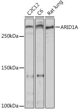
Figure 1. Western blot analysis of ARID1A using anti-ARID1A antibody (A00247-2). Electrophoresis was performed on a 5-20% SDS-PAGE gel at 70V (Stacking gel) / 90V (Resolving gel) for 2-3 hours. The sample well of each lane was loaded with 30 ug of sample under reducing conditions. Lane 1: human Hela whole cell lysates, Lane 2: human HepG2 whole cell lysates, Lane 3: human K562 whole cell lysates, Lane 4: human MCF-7 whole cell lysates. After electrophoresis, proteins were transferred to a nitrocellulose membrane at 150 mA for 50-90 minutes. Blocked the membrane with 5% non-fat milk/TBS for 1.5 hour at RT. The membrane was incubated with rabbit anti-ARID1A antigen affinity purified polyclonal antibody (Catalog # A00247-2) at 0.5 microg/mL overnight at 4°C, then washed with TBS-0.1%Tween 3 times with 5 minutes each and probed with a goat anti-rabbit IgG-HRP secondary antibody at a dilution of 1:5000 for 1.5 hour at RT. The signal is developed using an Enhanced Chemiluminescent detection (ECL) kit (Catalog # EK1002) with Tanon 5200 system. A specific band was detected for ARID1A at approximately 250-270 kDa. The expected band size for ARID1A is at 242 kDa.
. ARID1A was detected in a paraffin-embedded section of human colorectal adenocarcinoma tissue. Heat mediated antigen retrieval was performed in EDTA buffer (pH 8.0, epitope retrieval solution). The tissue section was blocked with 10% goat serum. The tissue section was then incubated with 2 microg/ml rabbit anti-ARID1A Antibody (A00247-2) overnight at 4°C. Peroxidase Conjugated Goat Anti-rabbit IgG was used as secondary antibody and incubated for 30 minutes at 37°C. The tissue section was developed using HRP Conjugated Rabbit IgG Super Vision Assay Kit (Catalog # SV0002) with DAB as the chromogen.")
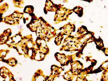
. ARID1A was detected in a paraffin-embedded section of human lung cancer tissue. Heat mediated antigen retrieval was performed in EDTA buffer (pH 8.0, epitope retrieval solution). The tissue section was blocked with 10% goat serum. The tissue section was then incubated with 2 microg/ml rabbit anti-ARID1A Antibody (A00247-2) overnight at 4°C. Peroxidase Conjugated Goat Anti-rabbit IgG was used as secondary antibody and incubated for 30 minutes at 37°C. The tissue section was developed using HRP Conjugated Rabbit IgG Super Vision Assay Kit (Catalog # SV0002) with DAB as the chromogen.")
. ARID1A was detected in a paraffin-embedded section of human spleen tissue. Heat mediated antigen retrieval was performed in EDTA buffer (pH 8.0, epitope retrieval solution). The tissue section was blocked with 10% goat serum. The tissue section was then incubated with 2 microg/ml rabbit anti-ARID1A Antibody (A00247-2) overnight at 4°C. Peroxidase Conjugated Goat Anti-rabbit IgG was used as secondary antibody and incubated for 30 minutes at 37°C. The tissue section was developed using HRP Conjugated Rabbit IgG Super Vision Assay Kit (Catalog # SV0002) with DAB as the chromogen.")
. ARID1A was detected in a paraffin-embedded section of rat thymus tissue. Heat mediated antigen retrieval was performed in EDTA buffer (pH 8.0, epitope retrieval solution). The tissue section was blocked with 10% goat serum. The tissue section was then incubated with 2 microg/ml rabbit anti-ARID1A Antibody (A00247-2) overnight at 4°C. Peroxidase Conjugated Goat Anti-rabbit IgG was used as secondary antibody and incubated for 30 minutes at 37°C. The tissue section was developed using HRP Conjugated Rabbit IgG Super Vision Assay Kit (Catalog # SV0002) with DAB as the chromogen.")
 and anti-Beta Tubulin antibody (M01857-3). ARID1A was detected in immunocytochemical section of A431 cell. Enzyme antigen retrieval was performed using IHC enzyme antigen retrieval reagent (AR0022) for 15 mins. The cells were blocked with 10% goat serum. And then incubated with 5 microg/mL rabbit anti-ARID1A Antibody (A00247-2) and mouse anti-Beta Tubulin antibody (M01857-3) overnight at 4°C. DyLight®488 Conjugated Goat Anti-Rabbit IgG (BA1127) and Cy3 Conjugated Goat Anti-Mouse IgG (BA1031) were used as secondary antibody at 1:500 dilution and incubated for 30 minutes at 37°C. Visualize using a fluorescence microscope and filter sets appropriate for the label used.")
. Overlay histogram showing K562 cells stained with A00247-2 (Blue line). To facilitate intracellular staining, cells were fixed with 4% paraformaldehyde and permeabilized with permeabilization buffer. The cells were blocked with 10% normal goat serum. And then incubated with rabbit anti-ARID1A Antibody (A00247-2, 1 microg/1x106 cells) for 30 min at 20°C. DyLight®488 conjugated goat anti-rabbit IgG (BA1127, 5-10 microg/1x106 cells) was used as secondary antibody for 30 minutes at 20°C. Isotype control antibody (Green line) was rabbit IgG (1 microg/1x106) used under the same conditions. Unlabelled sample (Red line) was also used as a control.")
Figure 1. Western blot analysis of ARID1A using anti-ARID1A antibody (A00247-2). Electrophoresis was performed on a 5-20% SDS-PAGE gel at 70V (Stacking gel) / 90V (Resolving gel) for 2-3 hours. The sample well of each lane was loaded with 30 ug of sample under reducing conditions. Lane 1: human Hela whole cell lysates, Lane 2: human HepG2 whole cell lysates, Lane 3: human K562 whole cell lysates, Lane 4: human MCF-7 whole cell lysates. After electrophoresis, proteins were transferred to a nitrocellulose membrane at 150 mA for 50-90 minutes. Blocked the membrane with 5% non-fat milk/TBS for 1.5 hour at RT. The membrane was incubated with rabbit anti-ARID1A antigen affinity purified polyclonal antibody (Catalog # A00247-2) at 0.5 microg/mL overnight at 4°C, then washed with TBS-0.1%Tween 3 times with 5 minutes each and probed with a goat anti-rabbit IgG-HRP secondary antibody at a dilution of 1:5000 for 1.5 hour at RT. The signal is developed using an Enhanced Chemiluminescent detection (ECL) kit (Catalog # EK1002) with Tanon 5200 system. A specific band was detected for ARID1A at approximately 250-270 kDa. The expected band size for ARID1A is at 242 kDa.
Anti-ARID1A Antibody Picoband(r)
A00247-2-CARRIER-FREE
ApplicationsFlow Cytometry, ImmunoFluorescence, Western Blot, ELISA, ImmunoCytoChemistry, ImmunoHistoChemistry
Product group Antibodies
ReactivityHuman, Rat
TargetARID1A
Overview
- SupplierBoster Bio
- Product NameAnti-ARID1A Antibody Picoband(r)
- Delivery Days Customer9
- ApplicationsFlow Cytometry, ImmunoFluorescence, Western Blot, ELISA, ImmunoCytoChemistry, ImmunoHistoChemistry
- CertificationResearch Use Only
- ClonalityPolyclonal
- Concentration500 ug/ml
- Gene ID8289
- Target nameARID1A
- Target descriptionAT-rich interaction domain 1A
- Target synonymsB120, BAF250, BAF250a, BM029, C1orf4, CSS2, ELD, MRD14, OSA1, P270, SMARCF1, hELD, hOSA1, AT-rich interactive domain-containing protein 1A, ARID domain-containing protein 1A, AT rich interactive domain 1A (SWI-like), BRG1-associated factor 250a, OSA1 nuclear protein, SWI-like protein, SWI/SNF complex protein p270, SWI/SNF-related, matrix-associated, actin-dependent regulator of chromatin subfamily F member 1, brain protein 120, chromatin remodeling factor p250, osa homolog 1
- HostRabbit
- IsotypeIgG
- Protein IDO14497
- Protein NameAT-rich interactive domain-containing protein 1A
- Scientific DescriptionBoster Bio Anti-ARID1A Antibody Picoband® catalog # A00247-2. Tested in ELISA, Flow Cytometry(Intracellular), IF, IHC, ICC, WB applications. This antibody reacts with Human, Rat. The brand Picoband indicates this is a premium antibody that guarantees superior quality, high affinity, and strong signals with minimal background in Western blot applications. Only our best-performing antibodies are designated as Picoband, ensuring unmatched performance.
- ReactivityHuman, Rat
- Storage Instruction-20°C,2°C to 8°C
- UNSPSC12352203
Related products
Product group Antibodies
Anti-ARID1A AntibodyA306631
ApplicationsWestern Blot
ReactivityHuman, Mouse, Rat
- SizePrice
Product group Antibodies
Anti-ARID1A (C-term) Antibody102-24767
ApplicationsWestern Blot
TargetARID1A
- SizePrice

Product group Antibodies
Anti-ARID1A AntibodyAMAB91192
ApplicationsWestern Blot, ImmunoCytoChemistry, ImmunoHistoChemistry
ReactivityHuman
TargetARID1A
- SizePrice
Product group Antibodies
ARID1A Recombinant Antibody, AbBy Fluor-350 ConjugatedBSM-61359R-BF350
ApplicationsImmunoFluorescence, Western Blot
ReactivityHuman, Mouse, Rat
TargetARID1A
- SizePrice
Product group Antibodies
ARID1A AntibodyCSB-PA002058LA01HU
ApplicationsELISA, ImmunoHistoChemistry
ReactivityHuman
TargetARID1A
- SizePrice
Product group Antibodies

ARID1A / BAF250 Antibody (aa95-108)LS-C402901
ApplicationsELISA, ImmunoHistoChemistry
ReactivityHuman, Mouse
TargetARID1A
- SizePrice
Product group Antibodies
ARID1A antibodyGTX129432
ApplicationsWestern Blot
ReactivityHuman
TargetARID1A
- SizePrice