

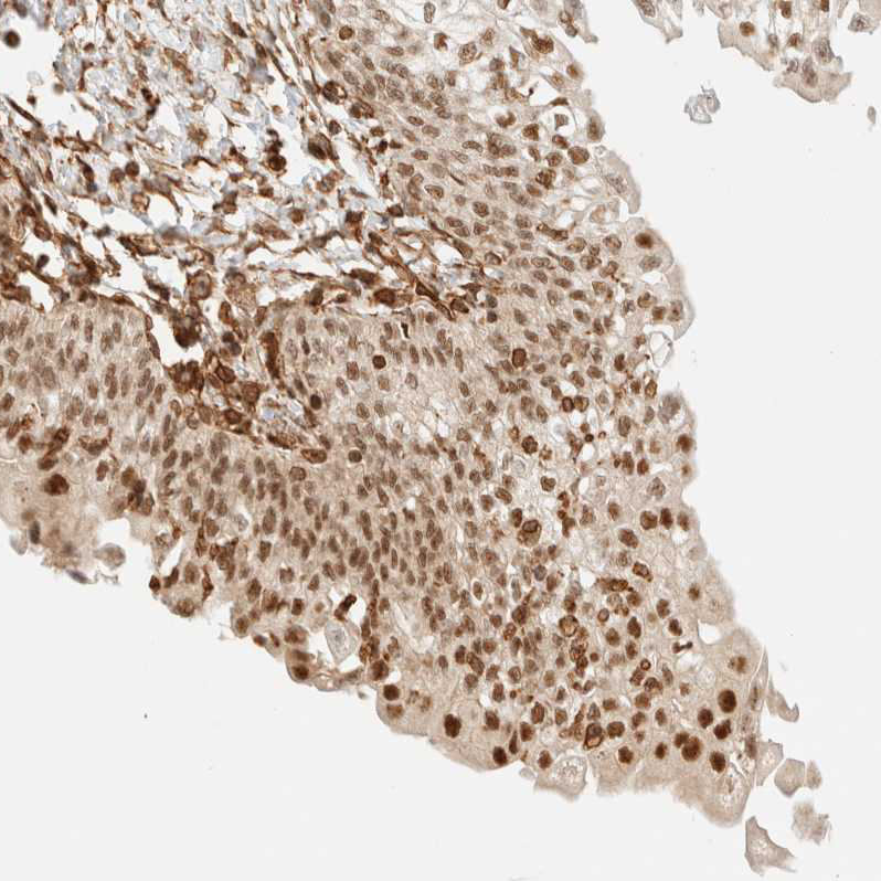
Immunohistochemical staining of human urinary bladder shows strong nuclear positivity in urothelial cells.

Immunohistochemical staining of human urinary bladder shows strong nuclear positivity in urothelial cells.
Anti-ARID5A Antibody
HPA023879
ApplicationsWestern Blot, ChIP Chromatin ImmunoPrecipitation, ImmunoCytoChemistry, ImmunoHistoChemistry
Product group Antibodies
ReactivityHuman
TargetARID5A
Overview
- SupplierAtlas Antibodies
- Product NameAnti-ARID5A Antibody
- Delivery Days Customer4
- ApplicationsWestern Blot, ChIP Chromatin ImmunoPrecipitation, ImmunoCytoChemistry, ImmunoHistoChemistry
- CertificationResearch Use Only
- ClonalityPolyclonal
- ConjugateUnconjugated
- Gene ID10865
- Target nameARID5A
- Target descriptionAT-rich interaction domain 5A
- Target synonymsMRF-1, MRF1, RP11-363D14, AT-rich interactive domain-containing protein 5A, ARID domain-containing protein 5A, AT rich interactive domain 5A (MRF1-like), RFVG5814, modulator recognition factor 1, modulator recognition factor I
- HostRabbit
- IsotypeIgG
- Protein IDQ03989
- Protein NameAT-rich interactive domain-containing protein 5A
- Scientific DescriptionRecombinant Protein Epitope Signature Tag (PrEST) antigen sequence
- ReactivityHuman
- Storage Instruction-20°C,2°C to 8°C
- UNSPSC41116161
Datasheet
MSDS
Related products
Product group Antibodies
Anti-ARID5A Antibody Picoband(r)A14678-1-CARRIER-FREE
ApplicationsWestern Blot, ELISA
ReactivityHuman
TargetARID5A
- SizePrice
Product group Antibodies

ARID5A Antibody (aa382-411)LS-C159114
ApplicationsWestern Blot
ReactivityHuman
TargetARID5A
- SizePrice
Product group Antibodies
ARID5A antibodyGTX122605
ApplicationsWestern Blot, ImmunoHistoChemistry, ImmunoHistoChemistry Paraffin
ReactivityHuman
TargetARID5A
- SizePrice
Product group Antibodies
Anti-ARID5A (Center) Antibody102-22837
ApplicationsWestern Blot
TargetARID5A
- SizePrice