


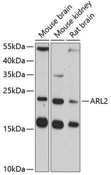
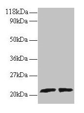
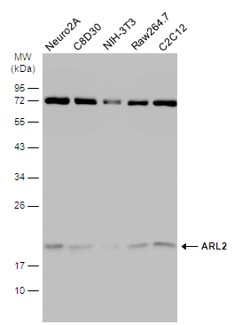
Figure 1. Western blot analysis of ARL2 using anti-ARL2 antibody (A04155-1). Electrophoresis was performed on a 5-20% SDS-PAGE gel at 70V (Stacking gel) / 90V (Resolving gel) for 2-3 hours. The sample well of each lane was loaded with 30 ug of sample under reducing conditions. Lane 1: human T-47D whole cell lysates, Lane 2: human A549 whole cell lysates, Lane 3: human U-397 whole cell lysates, Lane 4: human Hela whole cell lysates, Lane 5: rat liver tissue lysates, Lane 6: rat lung tissue lysates, Lane 7: mouse liver tissue lysates, Lane 8: mouse lung tissue lysates. After electrophoresis, proteins were transferred to a nitrocellulose membrane at 150 mA for 50-90 minutes. Blocked the membrane with 5% non-fat milk/TBS for 1.5 hour at RT. The membrane was incubated with rabbit anti-ARL2 antigen affinity purified polyclonal antibody (Catalog # A04155-1) at 0.5 microg/mL overnight at 4°C, then washed with TBS-0.1%Tween 3 times with 5 minutes each and probed with a goat anti-rabbit IgG-HRP secondary antibody at a dilution of 1:5000 for 1.5 hour at RT. The signal is developed using an Enhanced Chemiluminescent detection (ECL) kit (Catalog # EK1002) with Tanon 5200 system. A specific band was detected for ARL2 at approximately 18 kDa. The expected band size for ARL2 is at 21 kDa.
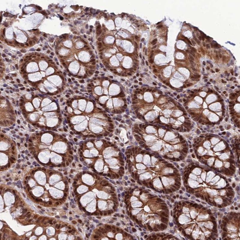
. ARL2 was detected in a paraffin-embedded section of human cervical cancer tissue. Heat mediated antigen retrieval was performed in EDTA buffer (pH 8.0, epitope retrieval solution). The tissue section was blocked with 10% goat serum. The tissue section was then incubated with 2 microg/ml rabbit anti-ARL2 Antibody (A04155-1) overnight at 4°C. Peroxidase Conjugated Goat Anti-rabbit IgG was used as secondary antibody and incubated for 30 minutes at 37°C. The tissue section was developed using HRP Conjugated Rabbit IgG Super Vision Assay Kit (Catalog # SV0002) with DAB as the chromogen.")
. ARL2 was detected in an immunocytochemical section of HELA cells. Enzyme antigen retrieval was performed using IHC enzyme antigen retrieval reagent (AR0022) for 15 mins. The cells were blocked with 10% goat serum. And then incubated with 5 microg/mL rabbit anti-ARL2 Antibody (A04155-1) overnight at 4°C. DyLight®488 Conjugated Goat Anti-Rabbit IgG (BA1127) was used as secondary antibody at 1:500 dilution and incubated for 30 minutes at 37°C. The section was counterstained with DAPI. Visualize using a fluorescence microscope and filter sets appropriate for the label used.")
. Overlay histogram showing JK cells stained with A04155-1 (Blue line). To facilitate intracellular staining, cells were fixed with 4% paraformaldehyde and permeabilized with permeabilization buffer. The cells were blocked with 10% normal goat serum. And then incubated with rabbit anti-ARL2 Antibody (A04155-1, 1 microg/1x106 cells) for 30 min at 20°C. DyLight®488 conjugated goat anti-rabbit IgG (BA1127, 5-10 microg/1x106 cells) was used as secondary antibody for 30 minutes at 20°C. Isotype control antibody (Green line) was rabbit IgG (1 microg/1x106) used under the same conditions. Unlabelled sample without incubation with primary antibody and secondary antibody (Red line) was used as a blank control.")
Figure 1. Western blot analysis of ARL2 using anti-ARL2 antibody (A04155-1). Electrophoresis was performed on a 5-20% SDS-PAGE gel at 70V (Stacking gel) / 90V (Resolving gel) for 2-3 hours. The sample well of each lane was loaded with 30 ug of sample under reducing conditions. Lane 1: human T-47D whole cell lysates, Lane 2: human A549 whole cell lysates, Lane 3: human U-397 whole cell lysates, Lane 4: human Hela whole cell lysates, Lane 5: rat liver tissue lysates, Lane 6: rat lung tissue lysates, Lane 7: mouse liver tissue lysates, Lane 8: mouse lung tissue lysates. After electrophoresis, proteins were transferred to a nitrocellulose membrane at 150 mA for 50-90 minutes. Blocked the membrane with 5% non-fat milk/TBS for 1.5 hour at RT. The membrane was incubated with rabbit anti-ARL2 antigen affinity purified polyclonal antibody (Catalog # A04155-1) at 0.5 microg/mL overnight at 4°C, then washed with TBS-0.1%Tween 3 times with 5 minutes each and probed with a goat anti-rabbit IgG-HRP secondary antibody at a dilution of 1:5000 for 1.5 hour at RT. The signal is developed using an Enhanced Chemiluminescent detection (ECL) kit (Catalog # EK1002) with Tanon 5200 system. A specific band was detected for ARL2 at approximately 18 kDa. The expected band size for ARL2 is at 21 kDa.
Anti-ARL2 Antibody Picoband(r)
A04155-1-CARRIER-FREE
ApplicationsFlow Cytometry, ImmunoFluorescence, Western Blot, ImmunoCytoChemistry, ImmunoHistoChemistry
Product group Antibodies
ReactivityHuman, Mouse, Rat
TargetARL2
Overview
- SupplierBoster Bio
- Product NameAnti-ARL2 Antibody Picoband(r)
- Delivery Days Customer9
- ApplicationsFlow Cytometry, ImmunoFluorescence, Western Blot, ImmunoCytoChemistry, ImmunoHistoChemistry
- CertificationResearch Use Only
- ClonalityPolyclonal
- Concentration500 ug/ml
- Gene ID402
- Target nameARL2
- Target descriptionARF like GTPase 2
- Target synonymsARFL2, MRCS1, ADP-ribosylation factor-like protein 2, ADP ribosylation factor like GTPase 2, ADP-ribosylation factor-like 2
- HostRabbit
- IsotypeIgG
- Protein IDP36404
- Protein NameADP-ribosylation factor-like protein 2
- Scientific DescriptionBoster Bio Anti-ARL2 Antibody Picoband® catalog # A04155-1. Tested in WB, IHC, ICC/IF, Flow Cytometry applications. This antibody reacts with Human, Mouse, Rat. The brand Picoband indicates this is a premium antibody that guarantees superior quality, high affinity, and strong signals with minimal background in Western blot applications. Only our best-performing antibodies are designated as Picoband, ensuring unmatched performance.
- ReactivityHuman, Mouse, Rat
- Storage Instruction-20°C,2°C to 8°C
- UNSPSC12352203
Related products
Product group Antibodies
Anti-ARL2 Antibody144-61352
ApplicationsWestern Blot
ReactivityHuman, Mouse, Rat
TargetARL2
- SizePrice
Product group Antibodies
Anti-ARL2 AntibodyA14214
ApplicationsWestern Blot
ReactivityMouse, Rat
- SizePrice
Product group Antibodies
ARL2 AntibodyCSB-PA01265A0RB
ApplicationsWestern Blot, ELISA
ReactivityHuman
TargetARL2
- SizePrice
Product group Antibodies

Goat anti-ARL2EB05545
ApplicationsFlow Cytometry, ImmunoFluorescence, Western Blot, ELISA
ReactivityCanine, Human
TargetARL2
- SizePrice
Product group Antibodies
ARL2 Polyclonal AntibodyCAC13797
ApplicationsWestern Blot, ELISA
TargetARL2
- SizePrice
Product group Antibodies

ARL2 AntibodyLS-C408559
ApplicationsWestern Blot
ReactivityHuman, Mouse, Rat
TargetARL2
- SizePrice
Product group Antibodies
ARL2 antibodyGTX100060
ApplicationsWestern Blot
ReactivityHuman, Mouse, Rat
TargetARL2
- SizePrice
Product group Antibodies
Anti-ARL2 AntibodyHPA044610
ApplicationsImmunoCytoChemistry, ImmunoHistoChemistry
ReactivityHuman
TargetARL2
- SizePrice
Product group Antibodies
Anti-ARL2 AntibodyCAB2830
ApplicationsWestern Blot, ELISA
ReactivityMouse
TargetARL2
- SizePrice