


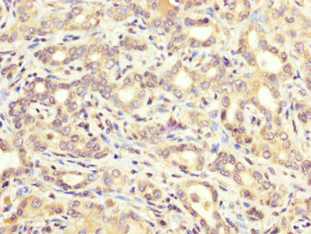
Immunohistochemical staining of human liver shows strong cytoplasmic positivity in hepatocytes.

Immunohistochemical staining of human liver shows strong cytoplasmic positivity in hepatocytes.
Anti-ATE1 Antibody
HPA038444
ApplicationsWestern Blot, ImmunoCytoChemistry, ImmunoHistoChemistry
Product group Antibodies
ReactivityHuman
TargetATE1
Overview
- SupplierAtlas Antibodies
- Product NameAnti-ATE1 Antibody
- Delivery Days Customer4
- ApplicationsWestern Blot, ImmunoCytoChemistry, ImmunoHistoChemistry
- CertificationResearch Use Only
- ClonalityPolyclonal
- ConjugateUnconjugated
- Gene ID11101
- Target nameATE1
- Target descriptionarginyltransferase 1
- Target synonymsarginyl-tRNA--protein transferase 1, R-transferase 1, arginine-tRNA--protein transferase 1, arginyl-tRNA-protein transferase
- HostRabbit
- IsotypeIgG
- Protein IDO95260
- Protein NameArginyl-tRNA--protein transferase 1
- Scientific DescriptionRecombinant Protein Epitope Signature Tag (PrEST) antigen sequence
- ReactivityHuman
- Storage Instruction-20°C,2°C to 8°C
- UNSPSC41116161
Datasheet
MSDS
Related products
Product group Antibodies
Anti-ATE1 AntibodyA28296
ApplicationsWestern Blot
ReactivityHuman, Mouse, Rat
- SizePrice

Product group Antibodies
Anti-ATE1 Antibody Picoband(r)A07459-1-CARRIER-FREE
ApplicationsWestern Blot, ELISA
ReactivityHuman
TargetATE1
- SizePrice
Product group Antibodies

ATE1 AntibodyLS-C831158
ApplicationsELISA, ImmunoHistoChemistry
ReactivityHuman, Mouse
TargetATE1
- SizePrice
Product group Antibodies
ATE1 AntibodyCSB-PA002268LA01HU
ApplicationsImmunoFluorescence, ELISA, ImmunoHistoChemistry
ReactivityHuman
TargetATE1
- SizePrice
Product group Antibodies
ATE1 Polyclonal AntibodyBS-7973R
ApplicationsImmunoFluorescence, Western Blot, ELISA, ImmunoCytoChemistry, ImmunoHistoChemistry, ImmunoHistoChemistry Frozen, ImmunoHistoChemistry Paraffin
ReactivityBovine, Canine, Equine, Human, Mouse, Porcine, Rabbit, Rat, Sheep
TargetATE1
- SizePrice

Product group Antibodies
Anti-ATE1 AntibodyHPA057549
ApplicationsImmunoCytoChemistry
ReactivityHuman
TargetATE1
- SizePrice
Product group Antibodies
ATE1 antibody [N1C1]GTX115577
ApplicationsWestern Blot, ImmunoHistoChemistry, ImmunoHistoChemistry Paraffin
ReactivityHuman
TargetATE1
- SizePrice