


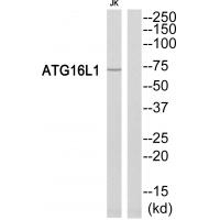
Figure 1. Western blot analysis of ATG16L1 using anti-ATG16L1 antibody (A00526). Electrophoresis was performed on a 5-20% SDS-PAGE gel at 70V (Stacking gel) / 90V (Resolving gel) for 2-3 hours. The sample well of each lane was loaded with 30 ug of sample under reducing conditions. Lane 1: human MCF-7 whole cell lysates, Lane 2: human Jurkat whole cell lysates, Lane 3: human 293T whole cell lysates, Lane 4: human PANC-1 whole cell lysates. After electrophoresis, proteins were transferred to a nitrocellulose membrane at 150 mA for 50-90 minutes. Blocked the membrane with 5% non-fat milk/TBS for 1.5 hour at RT. The membrane was incubated with rabbit anti-ATG16L1 antigen affinity purified polyclonal antibody (Catalog # A00526) at 0.5 microg/mL overnight at 4°C, then washed with TBS-0.1%Tween 3 times with 5 minutes each and probed with a goat anti-rabbit IgG-HRP secondary antibody at a dilution of 1:5000 for 1.5 hour at RT. The signal is developed using an Enhanced Chemiluminescent detection (ECL) kit (Catalog # EK1002) with Tanon 5200 system. A specific band was detected for ATG16L1 at approximately 68 kDa. The expected band size for ATG16L1 is at 68 kDa.
. ATG16L1 was detected in a paraffin-embedded section of human the renal pelvis is squamous metaplasia tissue. Heat mediated antigen retrieval was performed in EDTA buffer (pH 8.0, epitope retrieval solution). The tissue section was blocked with 10% goat serum. The tissue section was then incubated with 2 microg/ml rabbit anti-ATG16L1 Antibody (A00526-3) overnight at 4°C. Biotinylated goat anti-rabbit IgG was used as secondary antibody and incubated for 30 minutes at 37°C. The tissue section was developed using Strepavidin-Biotin-Complex (SABC) (Catalog # SA1022) with DAB as the chromogen.")
. ATG16L1 was detected in a paraffin-embedded section of human lung cancer tissue. Heat mediated antigen retrieval was performed in EDTA buffer (pH 8.0, epitope retrieval solution). The tissue section was blocked with 10% goat serum. The tissue section was then incubated with 2 microg/ml rabbit anti-ATG16L1 Antibody (A00526-3) overnight at 4°C. Biotinylated goat anti-rabbit IgG was used as secondary antibody and incubated for 30 minutes at 37°C. The tissue section was developed using Strepavidin-Biotin-Complex (SABC) (Catalog # SA1022) with DAB as the chromogen.")
. ATG16L1 was detected in a paraffin-embedded section of human esophageal squamous carcinoma tissue. Heat mediated antigen retrieval was performed in EDTA buffer (pH 8.0, epitope retrieval solution). The tissue section was blocked with 10% goat serum. The tissue section was then incubated with 2 microg/ml rabbit anti-ATG16L1 Antibody (A00526-3) overnight at 4°C. Biotinylated goat anti-rabbit IgG was used as secondary antibody and incubated for 30 minutes at 37°C. The tissue section was developed using Strepavidin-Biotin-Complex (SABC) (Catalog # SA1022) with DAB as the chromogen.")
. ATG16L1 was detected in an immunocytochemical section of A431 cells. Enzyme antigen retrieval was performed using IHC enzyme antigen retrieval reagent (AR0022) for 15 mins. The cells were blocked with 10% goat serum. And then incubated with 5 microg/mL rabbit anti-ATG16L1 Antibody (A00526-3) overnight at 4°C. DyLight®488 Conjugated Goat Anti-Rabbit IgG (BA1127) was used as secondary antibody at 1:100 dilution and incubated for 30 minutes at 37°C. The section was counterstained with DAPI. Visualize using a fluorescence microscope and filter sets appropriate for the label used.")
Figure 1. Western blot analysis of ATG16L1 using anti-ATG16L1 antibody (A00526). Electrophoresis was performed on a 5-20% SDS-PAGE gel at 70V (Stacking gel) / 90V (Resolving gel) for 2-3 hours. The sample well of each lane was loaded with 30 ug of sample under reducing conditions. Lane 1: human MCF-7 whole cell lysates, Lane 2: human Jurkat whole cell lysates, Lane 3: human 293T whole cell lysates, Lane 4: human PANC-1 whole cell lysates. After electrophoresis, proteins were transferred to a nitrocellulose membrane at 150 mA for 50-90 minutes. Blocked the membrane with 5% non-fat milk/TBS for 1.5 hour at RT. The membrane was incubated with rabbit anti-ATG16L1 antigen affinity purified polyclonal antibody (Catalog # A00526) at 0.5 microg/mL overnight at 4°C, then washed with TBS-0.1%Tween 3 times with 5 minutes each and probed with a goat anti-rabbit IgG-HRP secondary antibody at a dilution of 1:5000 for 1.5 hour at RT. The signal is developed using an Enhanced Chemiluminescent detection (ECL) kit (Catalog # EK1002) with Tanon 5200 system. A specific band was detected for ATG16L1 at approximately 68 kDa. The expected band size for ATG16L1 is at 68 kDa.
Anti-ATG16L1 Antibody Picoband(r)
A00526-3-CARRIER-FREE
ApplicationsImmunoFluorescence, Western Blot, ELISA, ImmunoCytoChemistry, ImmunoHistoChemistry
Product group Antibodies
ReactivityHuman
TargetATG16L1
Overview
- SupplierBoster Bio
- Product NameAnti-ATG16L1 Antibody Picoband(r)
- Delivery Days Customer9
- ApplicationsImmunoFluorescence, Western Blot, ELISA, ImmunoCytoChemistry, ImmunoHistoChemistry
- CertificationResearch Use Only
- ClonalityPolyclonal
- Concentration500 ug/ml
- Gene ID55054
- Target nameATG16L1
- Target descriptionautophagy related 16 like 1
- Target synonymsAPG16L, ATG16A, ATG16L, IBD10, WDR30, autophagy-related protein 16-1, APG16L beta, ATG16 autophagy related 16-like 1, WD repeat domain 30
- HostRabbit
- IsotypeIgG
- Scientific DescriptionBoster Bio Anti-ATG16L1 Antibody Picoband® catalog # A00526-3. Tested in ELISA, IF, IHC, ICC, WB applications. This antibody reacts with Human. The brand Picoband indicates this is a premium antibody that guarantees superior quality, high affinity, and strong signals with minimal background in Western blot applications. Only our best-performing antibodies are designated as Picoband, ensuring unmatched performance.
- ReactivityHuman
- Storage Instruction-20°C,2°C to 8°C
- UNSPSC12352203
Related products
Product group Antibodies
Anti-ATG16L1 Antibody144-60076
ApplicationsWestern Blot
ReactivityHuman
TargetATG16L1
- SizePrice
Product group Antibodies

ATG16L1 / ATG16L AntibodyLS-C831075
ApplicationsWestern Blot, ELISA, ImmunoHistoChemistry
ReactivityHuman, Mouse
TargetATG16L1
- SizePrice
Product group Antibodies
ATG16L1 Recombinant AntibodyBSM-60770R
ApplicationsWestern Blot
ReactivityHuman
TargetATG16L1
- SizePrice
Product group Antibodies

Goat anti-ATG16L1EB07893
ApplicationsWestern Blot, ELISA, ImmunoHistoChemistry
ReactivityCanine, Human, Mouse, Rat
TargetATG16L1
- SizePrice
Product group Antibodies
ApplicationsImmunoPrecipitation, Western Blot, ImmunoCytoChemistry, ImmunoHistoChemistry
ReactivityMouse, Porcine, Rat
TargetATG16L1
- SizePrice
Product group Antibodies
ATG16L1 AntibodyCSB-PA030171
ApplicationsWestern Blot, ELISA
ReactivityHuman
TargetATG16L1
- SizePrice
Product group Antibodies
Anti-ATG16L1 AntibodyHPA063900
ApplicationsImmunoHistoChemistry
ReactivityHuman
TargetATG16L1
- SizePrice
![Wild-type (WT) and ATG16L1 knockout (KO) HeLa cell extracts (30 μg) were separated by 7.5% SDS-PAGE, and the membrane was blotted with ATG16L1 antibody [N2C1], Internal (GTX110619) diluted at 1:2000. The HRP-conjugated anti-rabbit IgG antibody (GTX213110-01) was used to detect the primary antibody, and the signal was developed with Trident ECL plus-Enhanced.](https://www.genetex.com/upload/website/prouct_img/normal/GTX110619/GTX110619_40814_20180813_WB_KO_watermark_w_23060500_157.webp)
Product group Antibodies
ATG16L1 antibody [N2C1], InternalGTX110619
ApplicationsImmunoFluorescence, Western Blot, ImmunoCytoChemistry, ImmunoHistoChemistry, ImmunoHistoChemistry Paraffin
ReactivityHuman
TargetATG16L1
- SizePrice