


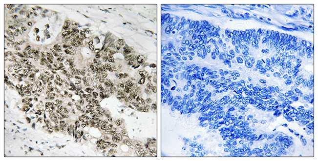
Figure 1. IF analysis of ATR using anti-ATR antibody (A00262-2). ATR was detected in an immunocytochemical section of A431 cells. Enzyme antigen retrieval was performed using IHC enzyme antigen retrieval reagent (AR0022) for 15 mins. The cells were blocked with 10% goat serum. And then incubated with 5 microg/mL rabbit anti-ATR Antibody (A00262-2) overnight at 4°C. DyLight®488 Conjugated Goat Anti-Rabbit IgG (BA1127) was used as secondary antibody at 1:100 dilution and incubated for 30 minutes at 37°C. The section was counterstained with DAPI. Visualize using a fluorescence microscope and filter sets appropriate for the label used.
Figure 1. IF analysis of ATR using anti-ATR antibody (A00262-2). ATR was detected in an immunocytochemical section of A431 cells. Enzyme antigen retrieval was performed using IHC enzyme antigen retrieval reagent (AR0022) for 15 mins. The cells were blocked with 10% goat serum. And then incubated with 5 microg/mL rabbit anti-ATR Antibody (A00262-2) overnight at 4°C. DyLight®488 Conjugated Goat Anti-Rabbit IgG (BA1127) was used as secondary antibody at 1:100 dilution and incubated for 30 minutes at 37°C. The section was counterstained with DAPI. Visualize using a fluorescence microscope and filter sets appropriate for the label used.
Anti-ATR Antibody
A00262-2-CARRIER-FREE
ApplicationsImmunoFluorescence, ELISA, ImmunoCytoChemistry
Product group Antibodies
ReactivityHuman
TargetATR
Overview
- SupplierBoster Bio
- Product NameAnti-ATR Antibody
- Delivery Days Customer9
- ApplicationsImmunoFluorescence, ELISA, ImmunoCytoChemistry
- CertificationResearch Use Only
- ClonalityPolyclonal
- Concentration500 ug/ml
- Gene ID545
- Target nameATR
- Target descriptionATR checkpoint kinase
- Target synonymsFCTCS, FRP1, MEC1, SCKL, SCKL1, serine/threonine-protein kinase ATR, ATR serine/threonine kinase, FRAP-related protein-1, MEC1, mitosis entry checkpoint 1, homolog, ataxia telangiectasia and Rad3-related protein
- HostRabbit
- IsotypeIgG
- Protein IDQ13535
- Protein NameSerine/threonine-protein kinase ATR
- Scientific DescriptionBoster Bio Anti-ATR Antibody Picoband® catalog # A00262-2. Tested in ELISA, IF, ICC applications. This antibody reacts with Human.
- ReactivityHuman
- Storage Instruction-20°C,2°C to 8°C
- UNSPSC12352203
Related products
Product group Antibodies
Anti-ATR AntibodyA101575
ApplicationsELISA, ImmunoHistoChemistry
ReactivityHuman
- SizePrice
Product group Antibodies
Anti-ATR Antibody144-63004
ApplicationsWestern Blot
ReactivityHuman, Mouse
TargetATR
- SizePrice
Product group Antibodies

ATR AntibodyLS-C832628
ApplicationsELISA, ImmunoHistoChemistry
ReactivityHuman
TargetATR
- SizePrice
Product group Antibodies
ATR Monoclonal AntibodyBSM-60260M
ApplicationsWestern Blot
ReactivityHuman
TargetATR
- SizePrice
Product group Antibodies
Atr Polyclonal AntibodyCAC09298
ApplicationsWestern Blot, ELISA, ImmunoHistoChemistry
TargetATR
- SizePrice
Product group Antibodies
Phospho-ATR (S428) AntibodyCSB-PA030127
ApplicationsELISA, ImmunoHistoChemistry
ReactivityHuman
TargetATR
- SizePrice

Product group Antibodies
Anti-ATR AntibodyHPA054320
ApplicationsImmunoCytoChemistry
ReactivityHuman
TargetATR
- SizePrice
Product group Antibodies
ATR (phospho Thr1989) antibodyGTX128145
ApplicationsImmunoFluorescence, ImmunoPrecipitation, Western Blot, ImmunoCytoChemistry, ImmunoHistoChemistry, ImmunoHistoChemistry Paraffin
ReactivityHuman, Mouse
TargetATR
- SizePrice