


Immunofluorescent staining of human cell line HeLa shows localization to nucleus.

Immunofluorescent staining of human cell line HeLa shows localization to nucleus.
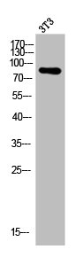
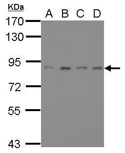
Anti-ATRIP Antibody
HPA047590
ApplicationsWestern Blot, ImmunoCytoChemistry
Product group Antibodies
ReactivityHuman
TargetATRIP
Overview
- SupplierAtlas Antibodies
- Product NameAnti-ATRIP Antibody
- Delivery Days Customer4
- ApplicationsWestern Blot, ImmunoCytoChemistry
- CertificationResearch Use Only
- ClonalityPolyclonal
- ConjugateUnconjugated
- Gene ID84126
- Target nameATRIP
- Target descriptionATR interacting protein
- Target synonymsATR-interacting protein, ATM and Rad3-related-interacting protein
- HostRabbit
- IsotypeIgG
- Protein IDQ8WXE1
- Protein NameATR-interacting protein
- Scientific DescriptionRecombinant Protein Epitope Signature Tag (PrEST) antigen sequence
- ReactivityHuman
- Storage Instruction-20°C,2°C to 8°C
- UNSPSC41116161
Datasheet
MSDS
Related products
Product group Antibodies
Anti-ATRIP AntibodyA101208
ApplicationsWestern Blot, ELISA
ReactivityHuman
- SizePrice

Product group Antibodies
Anti-ATRIP Antibody Picoband(r)A03862-2-CARRIER-FREE
ApplicationsFlow Cytometry, ImmunoFluorescence, Western Blot, ELISA, ImmunoCytoChemistry
ReactivityHuman, Monkey, Mouse, Rat
TargetATRIP
- SizePrice
Product group Antibodies
Anti-ATRIP Antibody144-07139
ApplicationsImmunoPrecipitation, Western Blot
ReactivityHuman, Mouse
TargetATRIP
- SizePrice
Product group Antibodies

ATRIP AntibodyLS-C830414
ApplicationsELISA, ImmunoHistoChemistry
ReactivityHuman, Mouse
TargetATRIP
- SizePrice
Product group Antibodies
ATRIP Recombinant Antibody, AbBy Fluor-647 ConjugatedBSM-62417R-BF647
ApplicationsWestern Blot
ReactivityHuman, Mouse, Rat
TargetATRIP
- SizePrice
Product group Antibodies
ATRIP AntibodyCSB-PA000958
ApplicationsImmunoFluorescence, Western Blot, ELISA, ImmunoHistoChemistry
ReactivityHuman, Mouse, Rat
TargetATRIP
- SizePrice
Product group Antibodies
ATRIP antibody [11]GTX70098
ApplicationsImmunoFluorescence, ImmunoPrecipitation, Western Blot, ImmunoCytoChemistry
ReactivityHuman
TargetATRIP
- SizePrice
Product group Antibodies
Anti-ATRIP AntibodyCAB5041
ApplicationsImmunoFluorescence, ImmunoPrecipitation, Western Blot, ELISA, ImmunoCytoChemistry
ReactivityHuman
TargetATRIP
- SizePrice