


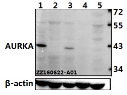
Figure 1. Western blot analysis of Aurora A/AURKA using anti-Aurora A/AURKA antibody (A00246-3). Electrophoresis was performed on a 5-20% SDS-PAGE gel at 70V (Stacking gel) / 90V (Resolving gel) for 2-3 hours. The sample well of each lane was loaded with 30 ug of sample under reducing conditions. Lane 1: human Hela whole cell lysates, Lane 2: human Caco-2 whole cell lysates, Lane 3: human HepG2 whole cell lysates, Lane 4: human SiHa whole cell lysates. After electrophoresis, proteins were transferred to a nitrocellulose membrane at 150 mA for 50-90 minutes. Blocked the membrane with 5% non-fat milk/TBS for 1.5 hour at RT. The membrane was incubated with rabbit anti-Aurora A/AURKA antigen affinity purified polyclonal antibody (Catalog # A00246-3) at 0.25 microg/mL overnight at 4°C, then washed with TBS-0.1%Tween 3 times with 5 minutes each and probed with a goat anti-rabbit IgG-HRP secondary antibody at a dilution of 1:5000 for 1.5 hour at RT. The signal is developed using an Enhanced Chemiluminescent detection (ECL) kit (Catalog # EK1002) with Tanon 5200 system. A specific band was detected for Aurora A/AURKA at approximately 50 kDa. The expected band size for Aurora A/AURKA is at 50 kDa.
. Aurora A/AURKA was detected in a paraffin-embedded section of human liver cancer tissue. Heat mediated antigen retrieval was performed in EDTA buffer (pH 8.0, epitope retrieval solution). The tissue section was blocked with 10% goat serum. The tissue section was then incubated with 2 microg/ml rabbit anti-Aurora A/AURKA Antibody (A00246-3) overnight at 4°C. Biotinylated goat anti-rabbit IgG was used as secondary antibody and incubated for 30 minutes at 37°C. The tissue section was developed using Strepavidin-Biotin-Complex (SABC) (Catalog # SA1022) with DAB as the chromogen.")
. Aurora A/AURKA was detected in a paraffin-embedded section of human Hashimoto thyroiditis tissue. Heat mediated antigen retrieval was performed in EDTA buffer (pH 8.0, epitope retrieval solution). The tissue section was blocked with 10% goat serum. The tissue section was then incubated with 2 microg/ml rabbit anti-Aurora A/AURKA Antibody (A00246-3) overnight at 4°C. Biotinylated goat anti-rabbit IgG was used as secondary antibody and incubated for 30 minutes at 37°C. The tissue section was developed using Strepavidin-Biotin-Complex (SABC) (Catalog # SA1022) with DAB as the chromogen.")
. Aurora A/AURKA was detected in an immunocytochemical section of Caco-2 cells. Enzyme antigen retrieval was performed using IHC enzyme antigen retrieval reagent (AR0022) for 15 mins. The cells were blocked with 10% goat serum. And then incubated with 5 microg/mL rabbit anti-Aurora A/AURKA Antibody (A00246-3) overnight at 4°C. DyLight®594 Conjugated Goat Anti-Rabbit IgG (BA1142) was used as secondary antibody at 1:100 dilution and incubated for 30 minutes at 37°C. The section was counterstained with DAPI. Visualize using a fluorescence microscope and filter sets appropriate for the label used.")
. Overlay histogram showing HL-60 cells stained with A00246-3 (Blue line). To facilitate intracellular staining, cells were fixed with 4% paraformaldehyde and permeabilized with permeabilization buffer. The cells were blocked with 10% normal goat serum. And then incubated with rabbit anti-Aurora A/AURKA Antibody (A00246-3, 1 microg/1x106 cells) for 30 min at 20°C. DyLight®488 conjugated goat anti-rabbit IgG (BA1127, 5-10 microg/1x106 cells) was used as secondary antibody for 30 minutes at 20°C. Isotype control antibody (Green line) was rabbit IgG (1 microg/1x106) used under the same conditions. Unlabelled sample without incubation with primary antibody and secondary antibody (Red line) was used as a blank control.")
Figure 1. Western blot analysis of Aurora A/AURKA using anti-Aurora A/AURKA antibody (A00246-3). Electrophoresis was performed on a 5-20% SDS-PAGE gel at 70V (Stacking gel) / 90V (Resolving gel) for 2-3 hours. The sample well of each lane was loaded with 30 ug of sample under reducing conditions. Lane 1: human Hela whole cell lysates, Lane 2: human Caco-2 whole cell lysates, Lane 3: human HepG2 whole cell lysates, Lane 4: human SiHa whole cell lysates. After electrophoresis, proteins were transferred to a nitrocellulose membrane at 150 mA for 50-90 minutes. Blocked the membrane with 5% non-fat milk/TBS for 1.5 hour at RT. The membrane was incubated with rabbit anti-Aurora A/AURKA antigen affinity purified polyclonal antibody (Catalog # A00246-3) at 0.25 microg/mL overnight at 4°C, then washed with TBS-0.1%Tween 3 times with 5 minutes each and probed with a goat anti-rabbit IgG-HRP secondary antibody at a dilution of 1:5000 for 1.5 hour at RT. The signal is developed using an Enhanced Chemiluminescent detection (ECL) kit (Catalog # EK1002) with Tanon 5200 system. A specific band was detected for Aurora A/AURKA at approximately 50 kDa. The expected band size for Aurora A/AURKA is at 50 kDa.
Anti-Aurora A/AURKA Antibody Picoband(r)
A00246-3-CARRIER-FREE
ApplicationsFlow Cytometry, ImmunoFluorescence, Western Blot, ELISA, ImmunoCytoChemistry, ImmunoHistoChemistry
Product group Antibodies
ReactivityHuman
TargetAURKA
Overview
- SupplierBoster Bio
- Product NameAnti-Aurora A/AURKA Antibody Picoband(r)
- Delivery Days Customer9
- ApplicationsFlow Cytometry, ImmunoFluorescence, Western Blot, ELISA, ImmunoCytoChemistry, ImmunoHistoChemistry
- CertificationResearch Use Only
- ClonalityPolyclonal
- Concentration500 ug/ml
- Gene ID6790
- Target nameAURKA
- Target descriptionaurora kinase A
- Target synonymsAIK, ARK1, AURA, BTAK, PPP1R47, STK15, STK6, STK7, aurora kinase A, aurora 2, aurora/IPL1-like kinase, aurora/IPL1-related kinase 1, breast tumor-amplified kinase, protein phosphatase 1, regulatory subunit 47, serine/threonine protein kinase 15, serine/threonine-protein kinase 6, serine/threonine-protein kinase aurora-A
- HostRabbit
- IsotypeIgG
- Protein IDO14965
- Protein NameAurora kinase A
- Scientific DescriptionBoster Bio Anti-Aurora A/AURKA Antibody Picoband® catalog # A00246-3. Tested in ELISA, Flow Cytometry, IF, IHC, ICC, WB applications. This antibody reacts with Human. The brand Picoband indicates this is a premium antibody that guarantees superior quality, high affinity, and strong signals with minimal background in Western blot applications. Only our best-performing antibodies are designated as Picoband, ensuring unmatched performance.
- ReactivityHuman
- Storage Instruction-20°C,2°C to 8°C
- UNSPSC12352203
Related products
Product group Antibodies
ApplicationsWestern Blot
ReactivityHuman, Mouse, Rat
- SizePrice
Product group Antibodies
Anti-AURKA Antibody144-02121
ApplicationsWestern Blot
ReactivityHuman, Mouse
TargetAURKA
- SizePrice
Product group Antibodies

AURKA / Aurora-A AntibodyLS-C831219
ApplicationsELISA, ImmunoHistoChemistry
ReactivityHuman
TargetAURKA
- SizePrice
Product group Antibodies
Aurora A Recombinant AntibodyBSM-52018R
ApplicationsImmunoFluorescence, ImmunoCytoChemistry, ImmunoHistoChemistry, ImmunoHistoChemistry Frozen, ImmunoHistoChemistry Paraffin
ReactivityHuman, Mouse, Rat
TargetAURKA
- SizePrice
Product group Antibodies
AURKA AntibodyCSB-PA000924
ApplicationsWestern Blot, ELISA
ReactivityHuman, Mouse, Rat
TargetAURKA
- SizePrice
Product group Antibodies
References

ApplicationsImmunoPrecipitation, Western Blot, ELISA
ReactivityHuman
TargetAURKA
- SizePrice
Product group Antibodies
Aurka Polyclonal AntibodyCAC07174
ApplicationsImmunoFluorescence, Western Blot, ELISA, ImmunoHistoChemistry
ReactivityMouse
TargetAURKA
- SizePrice

Product group Antibodies
Anti-AURKA AntibodyHPA002636
ApplicationsWestern Blot, ImmunoCytoChemistry, ImmunoHistoChemistry
ReactivityHuman
TargetAURKA
- SizePrice
![Aurora A antibody [C3], C-term detects Aurora A protein at spindle by immunofluorescent analysis. Sample: HeLa cells were fixed in ice-cold MeOH for 5 min. Green: Aurora A stained by Aurora A antibody [C3], C-term (GTX104620) diluted at 1:500. Red: alpha Tubulin, a cytoskeleton marker, stained by alpha Tubulin antibody [GT114] (GTX628802) diluted at 1:500. Blue: Hoechst 33342 staining.](https://www.genetex.com/upload/website/prouct_img/normal/GTX104620/GTX104620_43033_20180110_ICC_IF_w_23060120_926.webp)
Product group Antibodies
Aurora A antibody [C3], C-termGTX104620
ApplicationsImmunoFluorescence, Western Blot, ImmunoCytoChemistry, ImmunoHistoChemistry, ImmunoHistoChemistry Paraffin
ReactivityHuman
TargetAURKA
- SizePrice