


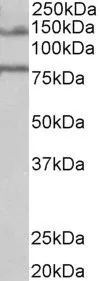
Figure 1. Western blot analysis of EPB41L5 using anti-EPB41L5 antibody (A09638-2). Electrophoresis was performed on a 5-20% SDS-PAGE gel at 70V (Stacking gel) / 90V (Resolving gel) for 2-3 hours. The sample well of each lane was loaded with 50ug of sample under reducing conditions. Lane 1: human HepG2 whole cell lysates, Lane 2: human HEK293 whole cell lysates. After Electrophoresis, proteins were transferred to a Nitrocellulose membrane at 150mA for 50-90 minutes. Blocked the membrane with 5% Non-fat Milk/ TBS for 1.5 hour at RT. The membrane was incubated with rabbit anti-EPB41L5 antigen affinity purified polyclonal antibody (Catalog # A09638-2) at 0.5 microg/mL overnight at 4°C, then washed with TBS-0.1%Tween 3 times with 5 minutes each and probed with a goat anti-rabbit IgG-HRP secondary antibody at a dilution of 1:5000 for 1.5 hour at RT. The signal is developed using an Enhanced Chemiluminescent detection (ECL) kit (Catalog # EK1002) with Tanon 5200 system. A specific band was detected for EPB41L5 at approximately 95KD. The expected band size for EPB41L5 is at 82KD.
. EPB41L5 was detected in paraffin-embedded section of human liver cancer tissue. Heat mediated antigen retrieval was performed in EDTA buffer (pH8.0, epitope retrieval solution). The tissue section was blocked with 10% goat serum. The tissue section was then incubated with 1microg/ml rabbit anti-EPB41L5 Antibody (A09638-2) overnight at 4°C. Biotinylated goat anti-rabbit IgG was used as secondary antibody and incubated for 30 minutes at 37°C. The tissue section was developed using Strepavidin-Biotin-Complex (SABC) (Catalog # SA1022) with DAB as the chromogen.")
. EPB41L5 was detected in paraffin-embedded section of human Ovarian cancer tissue. Heat mediated antigen retrieval was performed in EDTA buffer (pH8.0, epitope retrieval solution). The tissue section was blocked with 10% goat serum. The tissue section was then incubated with 1microg/ml rabbit anti-EPB41L5 Antibody (A09638-2) overnight at 4°C. Biotinylated goat anti-rabbit IgG was used as secondary antibody and incubated for 30 minutes at 37°C. The tissue section was developed using Strepavidin-Biotin-Complex (SABC) (Catalog # SA1022) with DAB as the chromogen.")
. Overlay histogram showing A431 cells stained with A09638-2 (Blue line). To facilitate intracellular staining, cells were fixed with 4% paraformaldehyde and permeabilized with permeabilization buffer. The cells were blocked with 10% normal goat serum. And then incubated with rabbit anti-EPB41L5 Antibody (A09638-2, 1microg/1x106 cells) for 30 min at 20°C. DyLight®488 conjugated goat anti-rabbit IgG (BA1127, 5-10microg/1x106 cells) was used as secondary antibody for 30 minutes at 20°C. Isotype control antibody (Green line) was rabbit IgG (1microg/1x106) used under the same conditions. Unlabelled sample without incubation with primary antibody and secondary antibody (Red line) was used as a blank control.")
Figure 1. Western blot analysis of EPB41L5 using anti-EPB41L5 antibody (A09638-2). Electrophoresis was performed on a 5-20% SDS-PAGE gel at 70V (Stacking gel) / 90V (Resolving gel) for 2-3 hours. The sample well of each lane was loaded with 50ug of sample under reducing conditions. Lane 1: human HepG2 whole cell lysates, Lane 2: human HEK293 whole cell lysates. After Electrophoresis, proteins were transferred to a Nitrocellulose membrane at 150mA for 50-90 minutes. Blocked the membrane with 5% Non-fat Milk/ TBS for 1.5 hour at RT. The membrane was incubated with rabbit anti-EPB41L5 antigen affinity purified polyclonal antibody (Catalog # A09638-2) at 0.5 microg/mL overnight at 4°C, then washed with TBS-0.1%Tween 3 times with 5 minutes each and probed with a goat anti-rabbit IgG-HRP secondary antibody at a dilution of 1:5000 for 1.5 hour at RT. The signal is developed using an Enhanced Chemiluminescent detection (ECL) kit (Catalog # EK1002) with Tanon 5200 system. A specific band was detected for EPB41L5 at approximately 95KD. The expected band size for EPB41L5 is at 82KD.
Anti-Band 4.1-like protein 5 EPB41L5 Antibody Picoband(r)
A09638-2-CARRIER-FREE
ApplicationsFlow Cytometry, Western Blot, ELISA, ImmunoHistoChemistry
Product group Antibodies
ReactivityHuman
TargetEPB41L5
Overview
- SupplierBoster Bio
- Product NameAnti-Band 4.1-like protein 5 EPB41L5 Antibody Picoband(r)
- Delivery Days Customer9
- ApplicationsFlow Cytometry, Western Blot, ELISA, ImmunoHistoChemistry
- CertificationResearch Use Only
- ClonalityPolyclonal
- Concentration500 ug/ml
- Gene ID57669
- Target nameEPB41L5
- Target descriptionerythrocyte membrane protein band 4.1 like 5
- Target synonymsBE37, LULU, LULU1, YMO1, YRT, band 4.1-like protein 5, yurt homolog
- HostRabbit
- IsotypeIgG
- Protein IDQ9HCM4
- Protein NameBand 4.1-like protein 5
- Scientific DescriptionBoster Bio Anti-Band 4.1-like protein 5 EPB41L5 Antibody catalog # A09638-2. Tested in ELISA, Flow Cytometry, IHC, WB applications. This antibody reacts with Human. The brand Picoband indicates this is a premium antibody that guarantees superior quality, high affinity, and strong signals with minimal background in Western blot applications. Only our best-performing antibodies are designated as Picoband, ensuring unmatched performance.
- ReactivityHuman
- Storage Instruction-20°C,2°C to 8°C
- UNSPSC12352203
Related products

Product group Antibodies

Goat anti-EBPL41L5EB08237
ApplicationsFlow Cytometry, ImmunoFluorescence, Western Blot, ELISA
ReactivityHuman
TargetEPB41L5
- SizePrice
Product group Antibodies
EBPL41L5 antibody, InternalGTX88887
ApplicationsWestern Blot
ReactivityHuman
TargetEPB41L5
- SizePrice
Product group Antibodies

EPB41L5 Antibody (aa355-383)LS-C159537
ApplicationsWestern Blot
ReactivityHuman
TargetEPB41L5
- SizePrice

Product group Antibodies
Anti-EPB41L5 AntibodyHPA037563
ApplicationsWestern Blot, ImmunoCytoChemistry, ImmunoHistoChemistry
ReactivityHuman
TargetEPB41L5
- SizePrice