


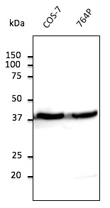
Figure 1. Western blot analysis of beta-Actin using anti-beta-Actin antibody (A01263-HRP). Electrophoresis was performed on a 5-20% SDS-PAGE gel at 70V (Stacking gel) / 90V (Resolving gel) for 2-3 hours. The sample well of each lane was loaded with 30 ug of sample under reducing conditions. Lane 1: human Hela whole cell lysates, Lane 2: human HepG2 whole cell lysates, Lane 3: human MCF-7 whole cell lysates, Lane 4: human K562 whole cell lysates, Lane 5: human A549 whole cell lysates, Lane 6: human A431 whole cell lysates, Lane 7: human U87 whole cell lysates, Lane 8: monkey COS-7 whole cell lysates. After electrophoresis, proteins were transferred to a nitrocellulose membrane at 150 mA for 50-90 minutes. Blocked the membrane with 5% non-fat milk/TBS for 1.5 hour at RT. The membrane was incubated with rabbit anti-beta-Actin antigen affinity purified polyclonal antibody (Catalog # A01263-HRP) at 0.1 microg/mL overnight at 4°C, then washed with TBS-0.1%Tween 3 times with 5 minutes each. The signal is developed using an Enhanced Chemiluminescent detection (ECL) kit (Catalog # EK1002) with Tanon 5200 system. A specific band was detected for beta-Actin at approximately 42 kDa. The expected band size for beta-Actin is at 42 kDa.
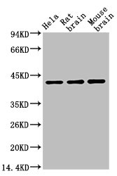
. Electrophoresis was performed on a 5-20% SDS-PAGE gel at 70V (Stacking gel) / 90V (Resolving gel) for 2-3 hours. The sample well of each lane was loaded with 30 ug of sample under reducing conditions. Lane 1: rat heart tissue lysates, Lane 2: rat skeletal muscle tissue lysates, Lane 3: rat brain tissue lysates, Lane 4: rat PC-12 whole cell lysates, Lane 5: mouse heart tissue lysates, Lane 6: mouse skeletal tissue lysates, Lane 7: mouse brain tissue lysates, Lane 8: mouse NIH/3T3 whole cell lysates. After electrophoresis, proteins were transferred to a nitrocellulose membrane at 150 mA for 50-90 minutes. Blocked the membrane with 5% non-fat milk/TBS for 1.5 hour at RT. The membrane was incubated with rabbit anti-beta-Actin antigen affinity purified polyclonal antibody (Catalog # A01263-HRP) at 0.1 microg/mL overnight at 4°C, then washed with TBS-0.1%Tween 3 times with 5 minutes each. The signal is developed using an Enhanced Chemiluminescent detection (ECL) kit (Catalog # EK1002) with Tanon 5200 system. A specific band was detected for beta-Actin at approximately 42 kDa. The expected band size for beta-Actin is at 42 kDa.")
Figure 1. Western blot analysis of beta-Actin using anti-beta-Actin antibody (A01263-HRP). Electrophoresis was performed on a 5-20% SDS-PAGE gel at 70V (Stacking gel) / 90V (Resolving gel) for 2-3 hours. The sample well of each lane was loaded with 30 ug of sample under reducing conditions. Lane 1: human Hela whole cell lysates, Lane 2: human HepG2 whole cell lysates, Lane 3: human MCF-7 whole cell lysates, Lane 4: human K562 whole cell lysates, Lane 5: human A549 whole cell lysates, Lane 6: human A431 whole cell lysates, Lane 7: human U87 whole cell lysates, Lane 8: monkey COS-7 whole cell lysates. After electrophoresis, proteins were transferred to a nitrocellulose membrane at 150 mA for 50-90 minutes. Blocked the membrane with 5% non-fat milk/TBS for 1.5 hour at RT. The membrane was incubated with rabbit anti-beta-Actin antigen affinity purified polyclonal antibody (Catalog # A01263-HRP) at 0.1 microg/mL overnight at 4°C, then washed with TBS-0.1%Tween 3 times with 5 minutes each. The signal is developed using an Enhanced Chemiluminescent detection (ECL) kit (Catalog # EK1002) with Tanon 5200 system. A specific band was detected for beta-Actin at approximately 42 kDa. The expected band size for beta-Actin is at 42 kDa.
Anti-beta-Actin ACTB Antibody Picoband(r) (HRP)
A01263-HRP
ApplicationsWestern Blot
Product group Antibodies
ReactivityChicken, Human, Monkey, Mouse, Rat, Zebra Fish
TargetACTB
Overview
- SupplierBoster Bio
- Product NameAnti-beta-Actin ACTB Antibody Picoband(r) (HRP)
- Delivery Days Customer9
- ApplicationsWestern Blot
- CertificationResearch Use Only
- ClonalityPolyclonal
- Concentration0.5 mg/ml
- ConjugateHRP
- Gene ID60
- Target nameACTB
- Target descriptionactin beta
- Target synonymsBKRNS, BNS, BRWS1, CSMH, DDS1, PS1TP5BP1, THC8, actin, cytoplasmic 1, I(2)-actin, PS1TP5-binding protein 1, beta cytoskeletal actin
- HostRabbit
- IsotypeIgG
- Protein IDP60709
- Protein NameActin, cytoplasmic 1
- Scientific DescriptionBoster Bio Anti-beta-Actin ACTB Antibody (HRP) (Catalog # A01263-HRP). Tested in WB applications. This antibody reacts with Human, Mouse, Rat, Monkey, Chicken, Zebrafish. The brand Picoband indicates this is a premium antibody that guarantees superior quality, high affinity, and strong signals with minimal background in Western blot applications. Only our best-performing antibodies are designated as Picoband, ensuring unmatched performance.
- ReactivityChicken, Human, Monkey, Mouse, Rat, Zebra Fish
- Storage Instruction-20°C,2°C to 8°C
- UNSPSC12352203
Related products
Product group Antibodies
Anti-beta Actin AntibodyA121645
ApplicationsImmunoFluorescence, Western Blot
ReactivityCanine, Human, Monkey, Mouse, Rat
- SizePrice
Product group Antibodies

Actin-pan AntibodyABX013019
ApplicationsWestern Blot, ELISA, ImmunoHistoChemistry
- SizePrice

Product group Antibodies
Anti-ACTB AntibodyAMAB91241
ApplicationsWestern Blot, ImmunoCytoChemistry, ImmunoHistoChemistry
ReactivityHuman, Mouse, Rat
TargetACTB
- SizePrice
Product group Antibodies

Anti-Beta-actin [Fab 19, actin]Ab02221-1.1
ApplicationsImmunoFluorescence, ELISA
ReactivityHuman
TargetACTB
- SizePrice
Product group Antibodies
ApplicationsWestern Blot, ELISA
ReactivityChicken, Hamster, Human, Monkey, Mouse, Rabbit, Rat, Sheep
TargetACTB
- SizePrice
Product group Antibodies

ACTB / Beta Actin AntibodyLS-C831486
ApplicationsWestern Blot, ImmunoHistoChemistry
ReactivityHuman, Mouse, Rat, Zebra Fish
TargetACTB
- SizePrice

Product group Antibodies
References
ApplicationsWestern Blot
ReactivityHuman, Mouse, Rat
TargetACTB
- SizePrice
Product group Antibodies
ACTB Monoclonal AntibodyCSB-MA000187
ApplicationsWestern Blot, ELISA
ReactivityCanine, Chicken, Hamster, Human, Monkey, Mouse, Porcine, Rabbit, Rat, Sheep
TargetACTB
- SizePrice
Product group Antibodies
Actb Polyclonal AntibodyCAC07011
ApplicationsImmunoFluorescence, Western Blot, ELISA, ImmunoHistoChemistry
TargetACTB
- SizePrice