


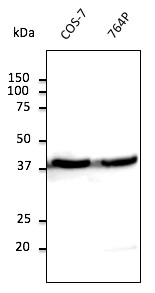
Figure 1. Western blot analysis of beta Actin/ACTB using anti-beta Actin/ACTB antibody (PA1872). Electrophoresis was performed on a 5-20% SDS-PAGE gel at 70V (Stacking gel) / 90V (Resolving gel) for 2-3 hours. The sample well of each lane was loaded with 30 ug of sample under reducing conditions. Lane 1: human Hela whole cell lysates, Lane 2: human K562 whole cell lysates, Lane 3: human MCF-7 whole cell lysates, Lane 4: human Jurkat whole cell lysates, Lane 5: rat brain tissue lysates, Lane 6: rat kidney tissue lysates, Lane 7: mouse brain tissue lysates, Lane 8: mouse kidney tissue lysates. After electrophoresis, proteins were transferred to a nitrocellulose membrane at 150 mA for 50-90 minutes. Blocked the membrane with 5% non-fat milk/TBS for 1.5 hour at RT. The membrane was incubated with rabbit anti-beta Actin/ACTB antigen affinity purified polyclonal antibody (Catalog # PA1872) at 0.5 microg/mL overnight at 4°C, then washed with TBS-0.1%Tween 3 times with 5 minutes each and probed with a goat anti-rabbit IgG-HRP secondary antibody at a dilution of 1:5000 for 1.5 hour at RT. The signal is developed using an Enhanced Chemiluminescent detection (ECL) kit (Catalog # EK1002) with Tanon 5200 system. A specific band was detected for beta Actin/ACTB at approximately 42 kDa. The expected band size for beta Actin/ACTB is at 42 kDa.
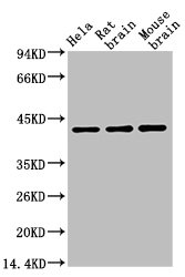
. Electrophoresis was performed on a 5-20% SDS-PAGE gel at 70V (Stacking gel) / 90V (Resolving gel) for 2-3 hours. The sample well of each lane was loaded with 30 ug of sample under reducing conditions. Lane 1: zebrafish tissue lysates, Lane 2: chicken heart tissue lysates, Lane 3: chicken liver tissue lysates, Lane 4: chicken brain tissue lysates, Lane 5: monkey heart tissue lysates, Lane 6: monkey lung tissue lysates, Lane 7: monkey kidney tissue lysates, Lane 8: monkey liver tissue lysates. After electrophoresis, proteins were transferred to a nitrocellulose membrane at 150 mA for 50-90 minutes. Blocked the membrane with 5% non-fat milk/TBS for 1.5 hour at RT. The membrane was incubated with rabbit anti-beta Actin/ACTB antigen affinity purified polyclonal antibody (Catalog # PA1872) at 0.5 microg/mL overnight at 4°C, then washed with TBS-0.1%Tween 3 times with 5 minutes each and probed with a goat anti-rabbit IgG-HRP secondary antibody at a dilution of 1:5000 for 1.5 hour at RT. The signal is developed using an Enhanced Chemiluminescent detection (ECL) kit (Catalog # EK1002) with Tanon 5200 system. A specific band was detected for beta Actin/ACTB at approximately 42 kDa. The expected band size for beta Actin/ACTB is at 42 kDa.")
Figure 1. Western blot analysis of beta Actin/ACTB using anti-beta Actin/ACTB antibody (PA1872). Electrophoresis was performed on a 5-20% SDS-PAGE gel at 70V (Stacking gel) / 90V (Resolving gel) for 2-3 hours. The sample well of each lane was loaded with 30 ug of sample under reducing conditions. Lane 1: human Hela whole cell lysates, Lane 2: human K562 whole cell lysates, Lane 3: human MCF-7 whole cell lysates, Lane 4: human Jurkat whole cell lysates, Lane 5: rat brain tissue lysates, Lane 6: rat kidney tissue lysates, Lane 7: mouse brain tissue lysates, Lane 8: mouse kidney tissue lysates. After electrophoresis, proteins were transferred to a nitrocellulose membrane at 150 mA for 50-90 minutes. Blocked the membrane with 5% non-fat milk/TBS for 1.5 hour at RT. The membrane was incubated with rabbit anti-beta Actin/ACTB antigen affinity purified polyclonal antibody (Catalog # PA1872) at 0.5 microg/mL overnight at 4°C, then washed with TBS-0.1%Tween 3 times with 5 minutes each and probed with a goat anti-rabbit IgG-HRP secondary antibody at a dilution of 1:5000 for 1.5 hour at RT. The signal is developed using an Enhanced Chemiluminescent detection (ECL) kit (Catalog # EK1002) with Tanon 5200 system. A specific band was detected for beta Actin/ACTB at approximately 42 kDa. The expected band size for beta Actin/ACTB is at 42 kDa.
Anti-beta Actin/ACTB Antibody Picoband(r)
PA1872
ApplicationsWestern Blot
Product group Antibodies
ReactivityChicken, Hamster, Human, Monkey, Mouse, Rat, Zebra Fish
TargetACTB
Overview
- SupplierBoster Bio
- Product NameAnti-beta Actin/ACTB Antibody Picoband(r)
- Delivery Days Customer9
- Application Supplier NoteTested Species: In-house tested species with positive results. Predicted Species: Species predicted to be fit for the product based on sequence similarities. Other applications have not been tested. Optimal dilutions should be determined by end users.
- ApplicationsWestern Blot
- Applications SupplierWB
- CertificationResearch Use Only
- ClonalityPolyclonal
- Concentration500 ug/ml
- Gene ID60
- Target nameACTB
- Target descriptionactin beta
- Target synonymsBKRNS, BNS, BRWS1, CSMH, DDS1, PS1TP5BP1, THC8, actin, cytoplasmic 1, I(2)-actin, PS1TP5-binding protein 1, beta cytoskeletal actin
- HostRabbit
- Protein IDP60709
- Protein NameActin, cytoplasmic 1
- Scientific DescriptionBoster Bio Anti-beta Actin/ACTB Antibody catalog # PA1872. Tested in WB applications. This antibody reacts with Human, Monkey, Mouse, Rat, Chicken, Zebrafish. The brand Picoband indicates this is a premium antibody that guarantees superior quality, high affinity, and strong signals with minimal background in Western blot applications. Only our best-performing antibodies are designated as Picoband, ensuring unmatched performance.
- ReactivityChicken, Hamster, Human, Monkey, Mouse, Rat, Zebra Fish
- Reactivity SupplierHuman, Mouse, Rat, Hamster
- Storage Instruction-20°C,2°C to 8°C
- UNSPSC12352203
References
- Shu L, Fu H, Pi A, et al. Protective effect of andrographolide against ulcerative colitis by activating Nrf2/HO-1 mediated antioxidant response. Front Pharmacol. 2024,15:1424219. doi: 10.3389/fphar.2024.1424219Read this paper
- Li F, Han X, Wu C, et al. Evaluation of immune and pyroptosis status in a model of sepsis-induced secondary pneumonia. Int Immunopharmacol. 2024,140:112835. doi: 10.1016/j.intimp.2024.112835Read this paper
- Li B, Chi X, Huang Y, et al. Bifidobacterium longum-Derived Extracellular Vesicles Prevent Hepatocellular Carcinoma by Modulating the TGF-β1/Smad Signaling in Mice. Front Biosci (Landmark Ed). 2024,29(7):241. doi: 10.31083/j.fbl2907241Read this paper
- Zeng LC, Zhang SH, Fu N, et al. miR-3202 inhibits bronchopulmonary dysplasia-mediated apoptosis and oxidative stress in bronchial epithelial cells via targeting RAG1. Pathol Res Pract. 2024,261:155482. doi: 10.1016/j.prp.2024.155482Read this paper
- Hao P, Li Q, Zhao H. Mucin 1 expression is regulated by hsa_circ_0055054/microRNA‑122‑5p and promotes hepatocellular carcinoma development. Oncol Lett. 2024,28(3):404. doi: 10.3892/ol.2024.14537Read this paper
- Yan X, Wang K, Shi C, et al. MicroRNA-138 promotes the progression of multiple myeloma through targeting paired PAX5. Mutat Res. 2024,829:111869. doi: 10.1016/j.mrfmmm.2024.111869Read this paper
- Meng X, Ren K, Liu X, et al. Efficacy of Rhamnus utilis Decne. Aqueous extract in mice with acute alcoholic liver injury and metabolomic study. Heliyon. 2024,10(12):e32523. doi: 10.1016/j.heliyon.2024.e32523Read this paper
- Pan L, She H, Hu Y, et al. Toll-like receptor 4 deficiency affects the balance of osteoclastogenesis and osteoblastogenesis in periodontitis. Int Immunopharmacol. 2024,137:112500. doi: 10.1016/j.intimp.2024.112500Read this paper
- Li X, Yuan F, Xiong Y, et al. FAM3A plays a key role in protecting against tubular cell pyroptosis and acute kidney injury. Redox Biol. 2024,74:103225. doi: 10.1016/j.redox.2024.103225Read this paper
- Li Y, Yang L, Su P, et al. Curcumin protects against cadmium-induced germ cell death in the testis of rats. Toxicol Res (Camb). 2024,13(2):tfae082. doi: 10.1093/toxres/tfae082Read this paper
Datasheet
MSDS
Related products
Product group Antibodies
Anti-beta Actin AntibodyA121645
ApplicationsImmunoFluorescence, Western Blot
ReactivityCanine, Human, Monkey, Mouse, Rat
- SizePrice
Product group Antibodies

Actin-pan AntibodyABX013019
ApplicationsWestern Blot, ELISA, ImmunoHistoChemistry
- SizePrice

Product group Antibodies
Anti-ACTB AntibodyAMAB91241
ApplicationsWestern Blot, ImmunoCytoChemistry, ImmunoHistoChemistry
ReactivityHuman, Mouse, Rat
TargetACTB
- SizePrice
Product group Antibodies

Anti-Beta-actin [Fab 19, actin]Ab02221-1.1
ApplicationsImmunoFluorescence, ELISA
ReactivityHuman
TargetACTB
- SizePrice
Product group Antibodies
ApplicationsWestern Blot, ELISA
ReactivityChicken, Hamster, Human, Monkey, Mouse, Rabbit, Rat, Sheep
TargetACTB
- SizePrice
Product group Antibodies

ACTB / Beta Actin AntibodyLS-C831486
ApplicationsWestern Blot, ImmunoHistoChemistry
ReactivityHuman, Mouse, Rat, Zebra Fish
TargetACTB
- SizePrice

Product group Antibodies
References
ApplicationsWestern Blot
ReactivityHuman, Mouse, Rat
TargetACTB
- SizePrice
Product group Antibodies
ACTB Monoclonal AntibodyCSB-MA000187
ApplicationsWestern Blot, ELISA
ReactivityCanine, Chicken, Hamster, Human, Monkey, Mouse, Porcine, Rabbit, Rat, Sheep
TargetACTB
- SizePrice
Product group Antibodies
Actb Polyclonal AntibodyCAC07011
ApplicationsImmunoFluorescence, Western Blot, ELISA, ImmunoHistoChemistry
TargetACTB
- SizePrice

Product group Antibodies
beta Actin antibodyGTX100313
ApplicationsImmunoFluorescence, Western Blot, ImmunoCytoChemistry, ImmunoHistoChemistry, ImmunoHistoChemistry Paraffin
ReactivityCanine, Drosophila, Feline, Human, Mouse
TargetACTB
- SizePrice