


Figure 7. Flow Cytometry analysis of HepG2 cells using anti-MAF antibody (A00654-1). Overlay histogram showing HepG2 cells stained with A00654-1 (Blue line). To facilitate intracellular staining, cells were fixed with 4% paraformaldehyde and permeabilized with permeabilization buffer. The cells were blocked with 10% normal goat serum. And then incubated with rabbit anti-MAF Antibody (A00654-1, 1microg/1x106 cells) for 30 min at 20°C. DyLight®488 conjugated goat anti-rabbit IgG (BA1127, 5-10microg/1x106 cells) was used as secondary antibody for 30 minutes at 20°C. Isotype control antibody (Green line) was rabbit IgG (1microg/1x106) used under the same conditions. Unlabelled sample without incubation with primary antibody and secondary antibody (Red line) was used as a blank control.
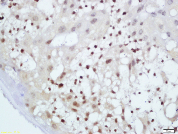
. MAF was detected in paraffin-embedded section of human tonsil tissue. Heat mediated antigen retrieval was performed in EDTA buffer (pH8.0, epitope retrieval solution). The tissue section was blocked with 10% goat serum. The tissue section was then incubated with 1microg/ml rabbit anti-MAF Antibody (A00654-1) overnight at 4°C. Biotinylated goat anti-rabbit IgG was used as secondary antibody and incubated for 30 minutes at 37°C. The tissue section was developed using Strepavidin-Biotin-Complex (SABC) (Catalog # SA1022) with DAB as the chromogen.")
. MAF was detected in paraffin-embedded section of human mammary cancer tissue. Heat mediated antigen retrieval was performed in EDTA buffer (pH8.0, epitope retrieval solution). The tissue section was blocked with 10% goat serum. The tissue section was then incubated with 1microg/ml rabbit anti-MAF Antibody (A00654-1) overnight at 4°C. Biotinylated goat anti-rabbit IgG was used as secondary antibody and incubated for 30 minutes at 37°C. The tissue section was developed using Strepavidin-Biotin-Complex (SABC) (Catalog # SA1022) with DAB as the chromogen.")
. MAF was detected in paraffin-embedded section of human placenta tissue. Heat mediated antigen retrieval was performed in EDTA buffer (pH8.0, epitope retrieval solution). The tissue section was blocked with 10% goat serum. The tissue section was then incubated with 1microg/ml rabbit anti-MAF Antibody (A00654-1) overnight at 4°C. Biotinylated goat anti-rabbit IgG was used as secondary antibody and incubated for 30 minutes at 37°C. The tissue section was developed using Strepavidin-Biotin-Complex (SABC) (Catalog # SA1022) with DAB as the chromogen.")
. MAF was detected in paraffin-embedded section of rat lung tissue. Heat mediated antigen retrieval was performed in EDTA buffer (pH8.0, epitope retrieval solution). The tissue section was blocked with 10% goat serum. The tissue section was then incubated with 1microg/ml rabbit anti-MAF Antibody (A00654-1) overnight at 4°C. Biotinylated goat anti-rabbit IgG was used as secondary antibody and incubated for 30 minutes at 37°C. The tissue section was developed using Strepavidin-Biotin-Complex (SABC) (Catalog # SA1022) with DAB as the chromogen.")
. MAF was detected in paraffin-embedded section of mouse liver tissue. Heat mediated antigen retrieval was performed in EDTA buffer (pH8.0, epitope retrieval solution). The tissue section was blocked with 10% goat serum. The tissue section was then incubated with 1microg/ml rabbit anti-MAF Antibody (A00654-1) overnight at 4°C. Biotinylated goat anti-rabbit IgG was used as secondary antibody and incubated for 30 minutes at 37°C. The tissue section was developed using Strepavidin-Biotin-Complex (SABC) (Catalog # SA1022) with DAB as the chromogen.")
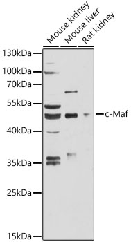
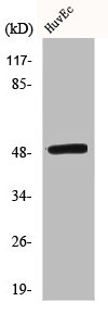
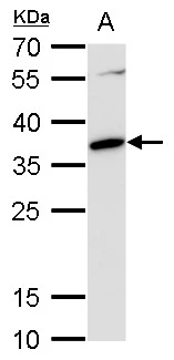
. Electrophoresis was performed on a 5-20% SDS-PAGE gel at 70V (Stacking gel) / 90V (Resolving gel) for 2-3 hours. The sample well of each lane was loaded with 50ug of sample under reducing conditions. Lane 1: human HEK293 whole cell lysates, Lane 2: human K562 whole cell lysates, Lane 3: human U2OS whole cell lysates, Lane 4: human placenta tissue lysates, Lane 5: human Caco-2 whole cell lysates, Lane 6: human PC-3 whole cell lysates, Lane 7: human HepG2 whole cell lysates, Lane 8: human THP-1 whole cell lysates. After Electrophoresis, proteins were transferred to a Nitrocellulose membrane at 150mA for 50-90 minutes. Blocked the membrane with 5% Non-fat Milk/ TBS for 1.5 hour at RT. The membrane was incubated with rabbit anti-MAF antigen affinity purified polyclonal antibody (Catalog # A00654-1) at 0.5 microg/mL overnight at 4°C, then washed with TBS-0.1%Tween 3 times with 5 minutes each and probed with a goat anti-rabbit IgG-HRP secondary antibody at a dilution of 1:10000 for 1.5 hour at RT. The signal is developed using an Enhanced Chemiluminescent detection (ECL) kit (Catalog # EK1002) with Tanon 5200 system. Specific bands were detected for MAF at approximately 42-50KD. The expected band size for MAF is at 38KD.")
Figure 7. Flow Cytometry analysis of HepG2 cells using anti-MAF antibody (A00654-1). Overlay histogram showing HepG2 cells stained with A00654-1 (Blue line). To facilitate intracellular staining, cells were fixed with 4% paraformaldehyde and permeabilized with permeabilization buffer. The cells were blocked with 10% normal goat serum. And then incubated with rabbit anti-MAF Antibody (A00654-1, 1microg/1x106 cells) for 30 min at 20°C. DyLight®488 conjugated goat anti-rabbit IgG (BA1127, 5-10microg/1x106 cells) was used as secondary antibody for 30 minutes at 20°C. Isotype control antibody (Green line) was rabbit IgG (1microg/1x106) used under the same conditions. Unlabelled sample without incubation with primary antibody and secondary antibody (Red line) was used as a blank control.
Anti-c-Maf/MAF Antibody Picoband(r)
A00654-1-CARRIER-FREE
ApplicationsFlow Cytometry, Western Blot, ImmunoHistoChemistry
Product group Antibodies
ReactivityHuman, Mouse, Rat
TargetMAF
Overview
- SupplierBoster Bio
- Product NameAnti-c-Maf/MAF Antibody Picoband(r)
- Delivery Days Customer9
- ApplicationsFlow Cytometry, Western Blot, ImmunoHistoChemistry
- CertificationResearch Use Only
- ClonalityPolyclonal
- Concentration500 ug/ml
- Gene ID4094
- Target nameMAF
- Target descriptionMAF bZIP transcription factor
- Target synonymsAYGRP, CCA4, CTRCT21, c-MAF, transcription factor Maf, Avian musculoaponeurotic fibrosarcoma (MAF) protooncogene, T lymphocyte c-maf long form, c-maf proto-oncogene, proto-oncogene c-Maf, v-maf avian musculoaponeurotic fibrosarcoma oncogene homolog
- HostRabbit
- IsotypeIgG
- Protein IDO75444
- Protein NameTranscription factor Maf
- Scientific DescriptionBoster Bio Anti-c-Maf/MAF Antibody Picoband® catalog # A00654-1. Tested in Flow Cytometry, IHC, WB applications. This antibody reacts with Human, Mouse, Rat. The brand Picoband indicates this is a premium antibody that guarantees superior quality, high affinity, and strong signals with minimal background in Western blot applications. Only our best-performing antibodies are designated as Picoband, ensuring unmatched performance.
- ReactivityHuman, Mouse, Rat
- Storage Instruction-20°C,2°C to 8°C
- UNSPSC12352203
Related products
Product group Antibodies
Anti-MAF Antibody144-62446
ApplicationsWestern Blot
ReactivityHuman, Mouse, Rat
TargetMAF
- SizePrice
Product group Antibodies
Anti-c-Maf AntibodyA89660
ApplicationsWestern Blot
ReactivityHuman, Mouse, Rat
- SizePrice
Product group Antibodies

c-Maf AntibodyLS-C831064
ApplicationsWestern Blot, ELISA, ImmunoHistoChemistry
ReactivityHuman, Mouse, Rat
TargetMAF
- SizePrice
Product group Antibodies
References
c-Maf Polyclonal AntibodyBS-5977R
ApplicationsImmunoFluorescence, ELISA, ImmunoCytoChemistry, ImmunoHistoChemistry, ImmunoHistoChemistry Frozen, ImmunoHistoChemistry Paraffin
ReactivityHuman, Mouse, Rat
TargetMAF
- SizePrice
Product group Antibodies
MAF AntibodyCSB-PA001726
ApplicationsWestern Blot, ELISA
ReactivityHuman, Mouse, Rat
TargetMAF
- SizePrice

Product group Antibodies
Anti-MAF AntibodyHPA028289
ApplicationsImmunoHistoChemistry
ReactivityHuman
TargetMAF
- SizePrice
Product group Antibodies
c-Maf antibodyGTX129420
ApplicationsWestern Blot, ChIP Chromatin ImmunoPrecipitation
ReactivityHuman
TargetMAF
- SizePrice