


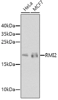
Figure 1. Western blot analysis of C16orf75/RMI2 using anti-C16orf75/RMI2 antibody (A08685-1). Electrophoresis was performed on a 5-20% SDS-PAGE gel at 70V (Stacking gel) / 90V (Resolving gel) for 2-3 hours. The sample well of each lane was loaded with 30 ug of sample under reducing conditions. Lane 1: human 293T whole cell lysates, Lane 2: human MCF-7 whole cell lysates, Lane 3: human HT-1080 whole cell lysates, Lane 4: human Hela whole cell lysates, Lane 5: rat thymus tissue lysates, Lane 6: mouse thymus tissue lysates. After electrophoresis, proteins were transferred to a nitrocellulose membrane at 150 mA for 50-90 minutes. Blocked the membrane with 5% non-fat milk/TBS for 1.5 hour at RT. The membrane was incubated with rabbit anti-C16orf75/RMI2 antigen affinity purified polyclonal antibody (Catalog # A08685-1) at 0.5 microg/mL overnight at 4°C, then washed with TBS-0.1%Tween 3 times with 5 minutes each and probed with a goat anti-rabbit IgG-HRP secondary antibody at a dilution of 1:5000 for 1.5 hour at RT. The signal is developed using an Enhanced Chemiluminescent detection (ECL) kit (Catalog # EK1002) with Tanon 5200 system. A specific band was detected for C16orf75/RMI2 at approximately 16 kDa. The expected band size for C16orf75/RMI2 is at 16 kDa.
Figure 1. Western blot analysis of C16orf75/RMI2 using anti-C16orf75/RMI2 antibody (A08685-1). Electrophoresis was performed on a 5-20% SDS-PAGE gel at 70V (Stacking gel) / 90V (Resolving gel) for 2-3 hours. The sample well of each lane was loaded with 30 ug of sample under reducing conditions. Lane 1: human 293T whole cell lysates, Lane 2: human MCF-7 whole cell lysates, Lane 3: human HT-1080 whole cell lysates, Lane 4: human Hela whole cell lysates, Lane 5: rat thymus tissue lysates, Lane 6: mouse thymus tissue lysates. After electrophoresis, proteins were transferred to a nitrocellulose membrane at 150 mA for 50-90 minutes. Blocked the membrane with 5% non-fat milk/TBS for 1.5 hour at RT. The membrane was incubated with rabbit anti-C16orf75/RMI2 antigen affinity purified polyclonal antibody (Catalog # A08685-1) at 0.5 microg/mL overnight at 4°C, then washed with TBS-0.1%Tween 3 times with 5 minutes each and probed with a goat anti-rabbit IgG-HRP secondary antibody at a dilution of 1:5000 for 1.5 hour at RT. The signal is developed using an Enhanced Chemiluminescent detection (ECL) kit (Catalog # EK1002) with Tanon 5200 system. A specific band was detected for C16orf75/RMI2 at approximately 16 kDa. The expected band size for C16orf75/RMI2 is at 16 kDa.
Anti-C16orf75/RMI2 Antibody Picoband(r)
A08685-1-CARRIER-FREE
ApplicationsWestern Blot, ELISA
Product group Antibodies
ReactivityHuman, Mouse, Rat
TargetRMI2
Overview
- SupplierBoster Bio
- Product NameAnti-C16orf75/RMI2 Antibody Picoband(r)
- Delivery Days Customer9
- ApplicationsWestern Blot, ELISA
- CertificationResearch Use Only
- ClonalityPolyclonal
- Concentration500 ug/ml
- Gene ID116028
- Target nameRMI2
- Target descriptionRecQ mediated genome instability 2
- Target synonymsBLAP18, C16orf75, recQ-mediated genome instability protein 2, BLM-associated protein of 18 kDa, RMI2, RecQ mediated genome instability 2, homolog
- HostRabbit
- IsotypeIgG
- Protein IDQ96E14
- Protein NameRecQ-mediated genome instability protein 2
- Scientific DescriptionBoster Bio Anti-C16orf75/RMI2 Antibody Picoband® catalog # A08685-1. Tested in ELISA, WB applications. This antibody reacts with Human, Mouse, Rat. The brand Picoband indicates this is a premium antibody that guarantees superior quality, high affinity, and strong signals with minimal background in Western blot applications. Only our best-performing antibodies are designated as Picoband, ensuring unmatched performance.
- ReactivityHuman, Mouse, Rat
- Storage Instruction-20°C,2°C to 8°C
- UNSPSC12352203
Related products
Product group Antibodies
Anti-RMI2 Antibody144-08523
ApplicationsWestern Blot
ReactivityHuman
TargetRMI2
- SizePrice

Product group Antibodies
RMI2 antibodyGTX65541
ApplicationsWestern Blot
ReactivityHuman
TargetRMI2
- SizePrice
Product group Antibodies
Anti-RMI2 AntibodyHPA040995
ApplicationsImmunoCytoChemistry, ImmunoHistoChemistry
ReactivityHuman
TargetRMI2
- SizePrice
Product group Antibodies

RMI2 / C16orf75 AntibodyLS-C410058
ApplicationsWestern Blot
ReactivityHuman
TargetRMI2
- SizePrice