


Immunofluorescent staining of human cell line U-251 MG shows localization to nucleoplasm.

Immunofluorescent staining of human cell line U-251 MG shows localization to nucleoplasm.
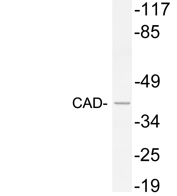
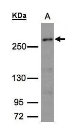
Anti-CAD Antibody
HPA057266
ApplicationsWestern Blot, ImmunoCytoChemistry
Product group Antibodies
ReactivityHuman
TargetCAD
Overview
- SupplierAtlas Antibodies
- Product NameAnti-CAD Antibody
- Delivery Days Customer4
- ApplicationsWestern Blot, ImmunoCytoChemistry
- CertificationResearch Use Only
- ClonalityPolyclonal
- ConjugateUnconjugated
- Gene ID790
- Target nameCAD
- Target descriptioncarbamoyl-phosphate synthetase 2, aspartate transcarbamylase, and dihydroorotase
- Target synonymsCDG1Z, DEE50, EIEE50, GATD4, multifunctional protein CAD, CAD trifunctional protein, carbamoyl phosphate synthetase 2-aspartate transcarbamylase-dihydroorotase
- HostRabbit
- IsotypeIgG
- Protein IDP27708
- Protein NameMultifunctional protein CAD
- Scientific DescriptionRecombinant Protein Epitope Signature Tag (PrEST) antigen sequence
- ReactivityHuman
- Storage Instruction-20°C,2°C to 8°C
- UNSPSC41116161
Datasheet
MSDS
Related products
Product group Antibodies
Anti-CAD AntibodyA101646
ApplicationsWestern Blot, ELISA
ReactivityHuman
- SizePrice
Product group Antibodies
Anti-CAD Antibody144-08344
ApplicationsWestern Blot, ImmunoHistoChemistry
ReactivityHuman
TargetCAD
- SizePrice
Product group Antibodies

CAD AntibodyLS-C829961
ApplicationsELISA, ImmunoHistoChemistry
ReactivityHuman, Mouse
TargetCAD
- SizePrice

Product group Antibodies
Anti-CAD/CAD Antibody Picoband(r)A00463-1-CARRIER-FREE
ApplicationsWestern Blot, ELISA
ReactivityHuman, Mouse
TargetCAD
- SizePrice
Product group Antibodies
CAD Recombinant Antibody, AbBy Fluor-405 ConjugatedBSM-62175R-BF405
ApplicationsFlow Cytometry, Western Blot
ReactivityHuman
TargetCAD
- SizePrice
Product group Antibodies

Goat anti-CAD / CPS2EB09418
ApplicationsELISA, ImmunoHistoChemistry
ReactivityHuman, Mouse, Rat
TargetCAD
- SizePrice
Product group Antibodies
Phospho-CAD (T456) AntibodyCSB-PA060131
ApplicationsELISA, ImmunoHistoChemistry
ReactivityHuman, Mouse
TargetCAD
- SizePrice
Product group Antibodies
CAD antibody [C2C3], C-termGTX101401
ApplicationsWestern Blot, ImmunoHistoChemistry, ImmunoHistoChemistry Paraffin
ReactivityHuman, Mouse
TargetCAD
- SizePrice