


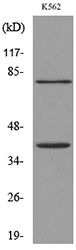
Figure 1. Western blot analysis of CCR2 using anti-CCR2 antibody (A00158-6). Electrophoresis was performed on a 5-20% SDS-PAGE gel at 70V (Stacking gel) / 90V (Resolving gel) for 2-3 hours. The sample well of each lane was loaded with 50ug of sample under reducing conditions. Lane 1: human HL-60 whole cell lysates, After Electrophoresis, proteins were transferred to a Nitrocellulose membrane at 150mA for 50-90 minutes. Blocked the membrane with 5% Non-fat Milk/ TBS for 1.5 hour at RT. The membrane was incubated with rabbit anti-CCR2 antigen affinity purified polyclonal antibody (Catalog # A00158-6) at 0.5 microg/mL overnight at 4°C, then washed with TBS-0.1%Tween 3 times with 5 minutes each and probed with a goat anti-rabbit IgG-HRP secondary antibody at a dilution of 1:5000 for 1.5 hour at RT. The signal is developed using an Enhanced Chemiluminescent detection (ECL) kit (Catalog # EK1002) with Tanon 5200 system. A specific band was detected for CCR2 at approximately 42KD. The expected band size for CCR2 is at 42KD.
. CCR2 was detected in paraffin-embedded section of human liver cancer tissue. Heat mediated antigen retrieval was performed in EDTA buffer (pH8.0, epitope retrieval solution). The tissue section was blocked with 10% goat serum. The tissue section was then incubated with 2microg/ml rabbit anti-CCR2 Antibody (A00158-6) overnight at 4°C. Biotinylated goat anti-rabbit IgG was used as secondary antibody and incubated for 30 minutes at 37°C. The tissue section was developed using Strepavidin-Biotin-Complex (SABC) (Catalog # SA1022) with DAB as the chromogen.")
. CCR2 was detected in paraffin-embedded section of human liver cancer tissue. Heat mediated antigen retrieval was performed in EDTA buffer (pH8.0, epitope retrieval solution). The tissue section was blocked with 10% goat serum. The tissue section was then incubated with 2microg/ml rabbit anti-CCR2 Antibody (A00158-6) overnight at 4°C. Biotinylated goat anti-rabbit IgG was used as secondary antibody and incubated for 30 minutes at 37°C. The tissue section was developed using Strepavidin-Biotin-Complex (SABC) (Catalog # SA1022) with DAB as the chromogen.")
. Overlay histogram showing h.PBMC cells stained with A00158-6 (Blue line).The cells were fixed with 4% paraformaldehyde and blocked with 10% normal goat serum. And then incubated with rabbit anti-CCR2 Antibody (A00158-6, 1microg/1x106 cells) for 30 min at 20°C. DyLight®488 conjugated goat anti-rabbit IgG (BA1127, 5-10microg/1x106 cells) was used as secondary antibody for 30 minutes at 20°C. Isotype control antibody (Green line) was rabbit IgG (1microg/1x106) used under the same conditions. Unlabelled sample without incubation with primary antibody and secondary antibody (Red line) was used as a blank control.")
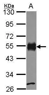
Figure 1. Western blot analysis of CCR2 using anti-CCR2 antibody (A00158-6). Electrophoresis was performed on a 5-20% SDS-PAGE gel at 70V (Stacking gel) / 90V (Resolving gel) for 2-3 hours. The sample well of each lane was loaded with 50ug of sample under reducing conditions. Lane 1: human HL-60 whole cell lysates, After Electrophoresis, proteins were transferred to a Nitrocellulose membrane at 150mA for 50-90 minutes. Blocked the membrane with 5% Non-fat Milk/ TBS for 1.5 hour at RT. The membrane was incubated with rabbit anti-CCR2 antigen affinity purified polyclonal antibody (Catalog # A00158-6) at 0.5 microg/mL overnight at 4°C, then washed with TBS-0.1%Tween 3 times with 5 minutes each and probed with a goat anti-rabbit IgG-HRP secondary antibody at a dilution of 1:5000 for 1.5 hour at RT. The signal is developed using an Enhanced Chemiluminescent detection (ECL) kit (Catalog # EK1002) with Tanon 5200 system. A specific band was detected for CCR2 at approximately 42KD. The expected band size for CCR2 is at 42KD.
Anti-CCR2 Antibody Picoband(r)
A00158-6-CARRIER-FREE
ApplicationsFlow Cytometry, Western Blot, ELISA, ImmunoHistoChemistry
Product group Antibodies
ReactivityHuman
TargetCD2
Overview
- SupplierBoster Bio
- Product NameAnti-CCR2 Antibody Picoband(r)
- Delivery Days Customer9
- ApplicationsFlow Cytometry, Western Blot, ELISA, ImmunoHistoChemistry
- CertificationResearch Use Only
- ClonalityPolyclonal
- Concentration500 ug/ml
- Gene ID914
- Target nameCD2
- Target descriptionCD2 molecule
- Target synonymsLFA-2, SRBC, T11, T-cell surface antigen CD2, CD2 antigen (p50), sheep red blood cell receptor, LFA-3 receptor, T-cell surface antigen T11/Leu-5, erythrocyte receptor, lymphocyte-function antigen-2, rosette receptor
- HostRabbit
- IsotypeIgG
- Protein IDP41597
- Protein NameC-C chemokine receptor type 2
- Scientific DescriptionBoster Bio Anti-CCR2 Antibody Picoband® catalog # A00158-6. Tested in ELISA, Flow Cytometry, IHC, WB applications. This antibody reacts with Human. The brand Picoband indicates this is a premium antibody that guarantees superior quality, high affinity, and strong signals with minimal background in Western blot applications. Only our best-performing antibodies are designated as Picoband, ensuring unmatched performance.
- ReactivityHuman
- Storage Instruction-20°C,2°C to 8°C
- UNSPSC12352203
Related products
Product group Antibodies

Anti-CD2 [YTH 655]Ab00164-10.0
ApplicationsFlow Cytometry, Other Application
ReactivityHuman
TargetCD2
- SizePrice

Product group Antibodies
Anti-CD2 AntibodyA101643
ApplicationsWestern Blot, ELISA
ReactivityHuman
- SizePrice
Product group Antibodies
Anti-CD2 Antibody144-65954
ApplicationsWestern Blot
ReactivityHuman, Mouse, Rat
TargetCD2
- SizePrice
Product group Antibodies

CD2 Antibody (clone TS1/8, FITC)LS-C812234
ApplicationsFlow Cytometry
ReactivityHuman
TargetCD2
- SizePrice
Product group Antibodies
CD2 Recombinant AntibodyBSM-60313R
ApplicationsImmunoFluorescence, ImmunoCytoChemistry, ImmunoHistoChemistry, ImmunoHistoChemistry Frozen, ImmunoHistoChemistry Paraffin
ReactivityHuman
TargetCD2
- SizePrice
Product group Antibodies
CD2 Monoclonal AntibodyCSB-MA000227
ApplicationsELISA, ImmunoHistoChemistry
ReactivityHuman, Mouse, Rat
TargetCD2
- SizePrice
Product group Antibodies
Cd2 Polyclonal AntibodyCAC07491
ApplicationsImmunoFluorescence, ELISA, ImmunoHistoChemistry
TargetCD2
- SizePrice
Product group Antibodies
CD2 antibody [C1C3]GTX101858
ApplicationsWestern Blot
ReactivityHuman
TargetCD2
- SizePrice

Product group Antibodies
Anti-CD2 AntibodyHPA003883
ApplicationsWestern Blot, ImmunoHistoChemistry
ReactivityHuman
TargetCD2
- SizePrice