

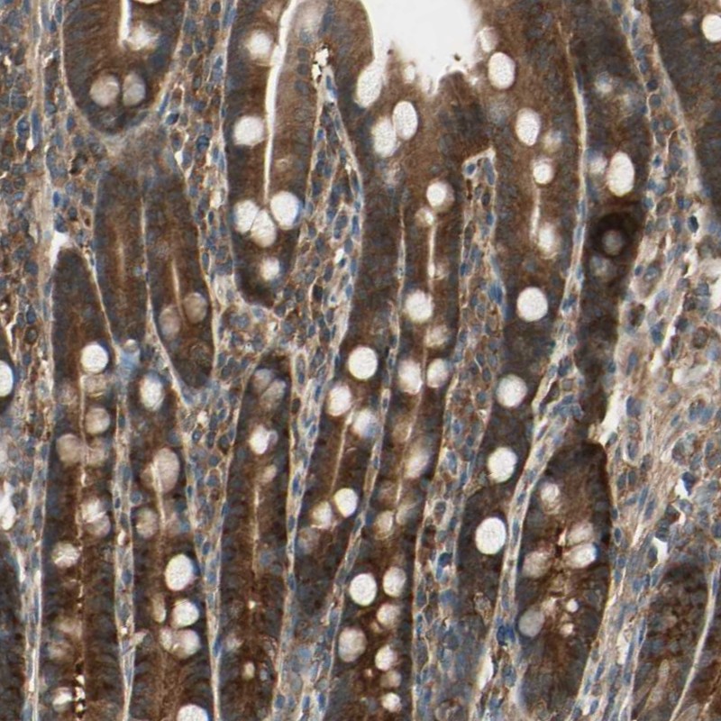
Immunohistochemical staining of human duodenum shows strong cytoplasmic positivity in glandular cells.

Immunohistochemical staining of human duodenum shows strong cytoplasmic positivity in glandular cells.
Anti-CCT7 Antibody
HPA008425
ApplicationsWestern Blot, ImmunoCytoChemistry, ImmunoHistoChemistry
Product group Antibodies
ReactivityHuman, Mouse, Rat
TargetCCT7
Overview
- SupplierAtlas Antibodies
- Product NameAnti-CCT7 Antibody
- Delivery Days Customer4
- ApplicationsWestern Blot, ImmunoCytoChemistry, ImmunoHistoChemistry
- CertificationResearch Use Only
- ClonalityPolyclonal
- ConjugateUnconjugated
- Gene ID10574
- Target nameCCT7
- Target descriptionchaperonin containing TCP1 subunit 7
- Target synonymsCCTETA, CCTH, NIP7-1, TCP1ETA, T-complex protein 1 subunit eta, CCT-eta, HIV-1 Nef interacting protein, TCP-1-eta, chaperonin containing T-complex polypeptide 1 subunit 7, chaperonin containing t-complex polypeptide 1, eta subunit
- HostRabbit
- IsotypeIgG
- Protein IDQ99832
- Protein NameT-complex protein 1 subunit eta
- Scientific DescriptionRecombinant Protein Epitope Signature Tag (PrEST) antigen sequence
- ReactivityHuman, Mouse, Rat
- Storage Instruction-20°C,2°C to 8°C
- UNSPSC41116161
Datasheet
MSDS
Related products
Product group Antibodies
Anti-CCT7 AntibodyA98250
ApplicationsWestern Blot, ELISA
ReactivityHuman, Mouse, Rat
- SizePrice

Product group Antibodies
Anti-TCP1 eta/CCT7 Antibody Picoband(r)A08169-2-CARRIER-FREE
ApplicationsFlow Cytometry, ImmunoFluorescence, Western Blot, ELISA, ImmunoCytoChemistry
ReactivityHuman, Mouse, Rat
TargetCCT7
- SizePrice
Product group Antibodies
Anti-CCT7 Antibody144-12146
ApplicationsWestern Blot
ReactivityHuman, Mouse, Rat
TargetCCT7
- SizePrice
Product group Antibodies

CCT7 AntibodyLS-C747321
ApplicationsWestern Blot
ReactivityHuman, Mouse, Rat
TargetCCT7
- SizePrice
Product group Antibodies
CCT7 AntibodyCSB-PA006639
ApplicationsWestern Blot, ELISA
ReactivityHuman, Mouse, Rat
TargetCCT7
- SizePrice
Product group Antibodies
CCT7 Polyclonal AntibodyCAC15146
ApplicationsImmunoFluorescence, Western Blot, ELISA, ImmunoHistoChemistry
TargetCCT7
- SizePrice
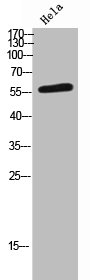
![Various whole cell extracts (30 μg) were separated by 7.5% SDS-PAGE, and the membrane was blotted with TCP1 eta antibody [C1C3] (GTX101347) diluted at 1:1000. The HRP-conjugated anti-rabbit IgG antibody (GTX213110-01) was used to detect the primary antibody.](https://www.genetex.com/upload/website/prouct_img/normal/GTX101347/GTX101347_40506_20211125_WB_w_23060100_690.webp)
Product group Antibodies
TCP1 eta antibody [C1C3]GTX101347
ApplicationsWestern Blot
ReactivityDrosophila, Human
TargetCCT7
- SizePrice