


Figure 1. Western blot analysis of CD326/Epcam using anti-CD326/Epcam antibody (A00276-2). Electrophoresis was performed on a 5-20% SDS-PAGE gel at 70V (Stacking gel) / 90V (Resolving gel) for 2-3 hours. The sample well of each lane was loaded with 30 ug of sample under reducing conditions. Lane 1: mouse small intestine tissue lysates, Lane 2: mouse kidney tissue lysates. After electrophoresis, proteins were transferred to a nitrocellulose membrane at 150 mA for 50-90 minutes. Blocked the membrane with 5% non-fat milk/TBS for 1.5 hour at RT. The membrane was incubated with rabbit anti-CD326/Epcam antigen affinity purified polyclonal antibody (Catalog # A00276-2) at 0.25 microg/mL overnight at 4°C, then washed with TBS-0.1%Tween 3 times with 5 minutes each and probed with a goat anti-rabbit IgG-HRP secondary antibody at a dilution of 1:5000 for 1.5 hour at RT. The signal is developed using an Enhanced Chemiluminescent detection (ECL) kit (Catalog # EK1002) with Tanon 5200 system. A specific band was detected for CD326/Epcam at approximately 38 kDa. The expected band size for CD326/Epcam is at 35,40 kDa.
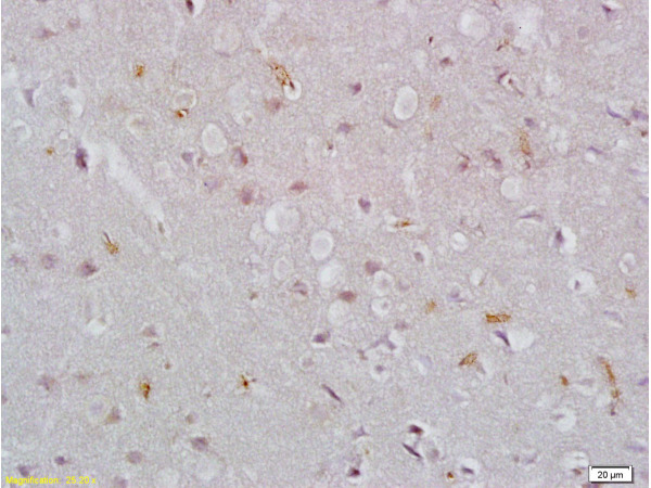
. CD326/Epcam was detected in a paraffin-embedded section of rat colon tissue. Heat mediated antigen retrieval was performed in EDTA buffer (pH 8.0, epitope retrieval solution). The tissue section was blocked with 10% goat serum. The tissue section was then incubated with 2 microg/ml rabbit anti-CD326/Epcam Antibody (A00276-2) overnight at 4°C. Peroxidase Conjugated Goat Anti-rabbit IgG was used as secondary antibody and incubated for 30 minutes at 37°C. The tissue section was developed using HRP Conjugated Rabbit IgG Super Vision Assay Kit (Catalog # SV0002) with DAB as the chromogen.")
. CD326/Epcam was detected in a paraffin-embedded section of mouse colon tissue. Heat mediated antigen retrieval was performed in EDTA buffer (pH 8.0, epitope retrieval solution). The tissue section was blocked with 10% goat serum. The tissue section was then incubated with 2 microg/ml rabbit anti-CD326/Epcam Antibody (A00276-2) overnight at 4°C. Peroxidase Conjugated Goat Anti-rabbit IgG was used as secondary antibody and incubated for 30 minutes at 37°C. The tissue section was developed using HRP Conjugated Rabbit IgG Super Vision Assay Kit (Catalog # SV0002) with DAB as the chromogen.")
. CD326/Epcam was detected in a paraffin-embedded section of mouse colon cancer tissue. Heat mediated antigen retrieval was performed in EDTA buffer (pH 8.0, epitope retrieval solution). The tissue section was blocked with 10% goat serum. The tissue section was then incubated with 5 microg/mL rabbit anti-CD326/Epcam Antibody (A00276-2) overnight at 4°C. FITC Conjugated Goat Anti-Rabbit IgG (BA1105) was used as secondary antibody at 1:500 dilution and incubated for 30 minutes at 37°C. The section was counterstained with DAPI. Visualize using a fluorescence microscope and filter sets appropriate for the label used.")
. CD326/Epcam was detected in a paraffin-embedded section of rat colon cancer tissue. Heat mediated antigen retrieval was performed in EDTA buffer (pH 8.0, epitope retrieval solution). The tissue section was blocked with 10% goat serum. The tissue section was then incubated with 5 microg/mL rabbit anti-CD326/Epcam Antibody (A00276-2) overnight at 4°C. FITC Conjugated Goat Anti-Rabbit IgG (BA1105) was used as secondary antibody at 1:500 dilution and incubated for 30 minutes at 37°C. The section was counterstained with DAPI. Visualize using a fluorescence microscope and filter sets appropriate for the label used.")
. Overlay histogram showing RAW264.7 cells stained with A00276-2 (Blue line). The cells were fixed with 4% paraformaldehyde and blocked with 10% normal goat serum. And then incubated with rabbit anti-CD326/Epcam Antibody (A00276-2, 1 microg/1x106 cells) for 30 min at 20°C. DyLight®488 conjugated goat anti-rabbit IgG (BA1127, 5-10 microg/1x106 cells) was used as secondary antibody for 30 minutes at 20°C. Isotype control antibody (Green line) was rabbit IgG (1 microg/1x106) used under the same conditions. Unlabelled sample (Red line) was also used as a control.")
Figure 1. Western blot analysis of CD326/Epcam using anti-CD326/Epcam antibody (A00276-2). Electrophoresis was performed on a 5-20% SDS-PAGE gel at 70V (Stacking gel) / 90V (Resolving gel) for 2-3 hours. The sample well of each lane was loaded with 30 ug of sample under reducing conditions. Lane 1: mouse small intestine tissue lysates, Lane 2: mouse kidney tissue lysates. After electrophoresis, proteins were transferred to a nitrocellulose membrane at 150 mA for 50-90 minutes. Blocked the membrane with 5% non-fat milk/TBS for 1.5 hour at RT. The membrane was incubated with rabbit anti-CD326/Epcam antigen affinity purified polyclonal antibody (Catalog # A00276-2) at 0.25 microg/mL overnight at 4°C, then washed with TBS-0.1%Tween 3 times with 5 minutes each and probed with a goat anti-rabbit IgG-HRP secondary antibody at a dilution of 1:5000 for 1.5 hour at RT. The signal is developed using an Enhanced Chemiluminescent detection (ECL) kit (Catalog # EK1002) with Tanon 5200 system. A specific band was detected for CD326/Epcam at approximately 38 kDa. The expected band size for CD326/Epcam is at 35,40 kDa.
Anti-CD326/Epcam Antibody Picoband(r)
A00276-2-PE
ApplicationsFlow Cytometry, ImmunoFluorescence, Western Blot, ELISA, ImmunoHistoChemistry
Product group Antibodies
ReactivityMouse, Rat
TargetEpcam
Overview
- SupplierBoster Bio
- Product NameAnti-CD326/Epcam Antibody Picoband(r)
- Delivery Days Customer9
- ApplicationsFlow Cytometry, ImmunoFluorescence, Western Blot, ELISA, ImmunoHistoChemistry
- CertificationResearch Use Only
- ClonalityPolyclonal
- Concentration500 ug/ml
- ConjugateRPE
- Gene ID17075
- Target nameEpcam
- Target descriptionepithelial cell adhesion molecule
- Target synonymsCD326, EGP, EGP-2, Egp314, Ep-CAM, EpCAM1, GA733-2, Ly74, TROP1, Tacsd1, Tacstd1, gp40, epithelial cell adhesion molecule, Trop-1 protein, epithelial glycoprotein 314, lymphocyte antigen 74, mEGP314, panepithelial glycoprotein 314, protein 289A, tumor-associated calcium signal transducer 1
- HostRabbit
- IsotypeIgG
- Protein IDQ99JW5
- Protein NameEpithelial cell adhesion molecule
- Scientific DescriptionBoster Bio Anti-CD326/Epcam Antibody Picoband® catalog # A00276-2. Tested in ELISA, Flow Cytometry, IF, IHC, WB applications. This antibody reacts with Mouse, Rat. The brand Picoband indicates this is a premium antibody that guarantees superior quality, high affinity, and strong signals with minimal background in Western blot applications. Only our best-performing antibodies are designated as Picoband, ensuring unmatched performance.
- ReactivityMouse, Rat
- Storage Instruction-20°C,2°C to 8°C
- UNSPSC12352203
Related products

Product group Antibodies
Anti-EpCAM AntibodyA00276-1-CARRIER-FREE
ApplicationsImmunoFluorescence, ELISA, ImmunoHistoChemistry
ReactivityMouse, Rat
TargetEpcam
- SizePrice
Product group Antibodies
ApplicationsFlow Cytometry
ReactivityMouse
TargetEpcam
- SizePrice
Product group Antibodies
Anti-Mouse Epcam (C-term) Antibody102-24235
ApplicationsFlow Cytometry, Western Blot
TargetEpcam
- SizePrice
Product group Antibodies
EpCAM Polyclonal AntibodyBS-0593R
ApplicationsImmunoFluorescence, Western Blot, ELISA, ImmunoCytoChemistry, ImmunoHistoChemistry, ImmunoHistoChemistry Frozen, ImmunoHistoChemistry Paraffin
ReactivityBovine, Canine, Human, Mouse, Porcine, Rabbit, Rat
TargetEpcam
- SizePrice
Product group Antibodies
Epcam AntibodyCSB-PA007717ZA01MO
ApplicationsWestern Blot, ELISA
ReactivityMouse
TargetEpcam
- SizePrice
Product group Antibodies

Rat anti EpCAM / CD326MUB0509P
ApplicationsFlow Cytometry, ImmunoPrecipitation, Western Blot, ImmunoCytoChemistry, ImmunoHistoChemistry, ImmunoHistoChemistry Frozen
ReactivityMouse
TargetEpcam
- SizePrice