


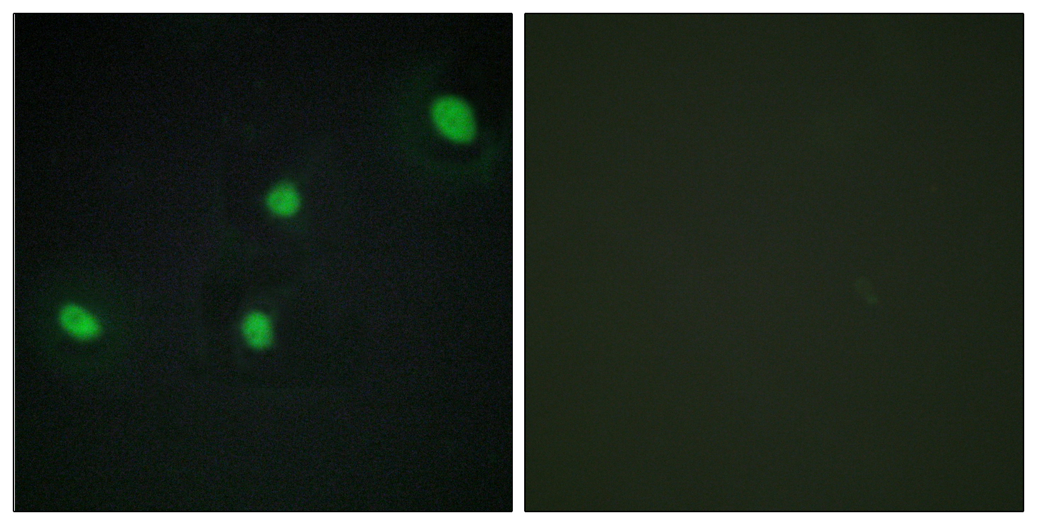
Immunohistochemistry analysis of paraffin-embedded human brain tissue, using CDCA4 Antibody. The picture on the right is blocked with the synthesized peptide.

Immunohistochemistry analysis of paraffin-embedded human brain tissue, using CDCA4 Antibody. The picture on the right is blocked with the synthesized peptide.
Anti-CDCA4 Antibody
A13717-1
ApplicationsImmunoFluorescence, Western Blot, ELISA, ImmunoCytoChemistry, ImmunoHistoChemistry
Product group Antibodies
ReactivityHuman, Mouse
TargetCDCA4
Overview
- SupplierBoster Bio
- Product NameAnti-CDCA4 Antibody
- Delivery Days Customer9
- ApplicationsImmunoFluorescence, Western Blot, ELISA, ImmunoCytoChemistry, ImmunoHistoChemistry
- CertificationResearch Use Only
- ClonalityPolyclonal
- Concentration0.5-1 mg/ml
- Gene ID55038
- Target nameCDCA4
- Target descriptioncell division cycle associated 4
- Target synonymsHEPP, SEI-3/HEPP, cell division cycle-associated protein 4, hematopoietic progenitor protein
- HostRabbit
- IsotypeIgG
- Protein IDQ9BXL8
- Protein NameCell division cycle-associated protein 4
- Scientific DescriptionBoster Bio Anti-CDCA4 Antibody (Catalog# A13717-1). Tested in IHC, ICC, IF, WB, ELISA applications. This antibody reacts with Human, Mouse.
- ReactivityHuman, Mouse
- Storage Instruction-20°C,2°C to 8°C
- UNSPSC12352203
Related products
Product group Antibodies
Anti-CDCA4 AntibodyA97651
ApplicationsImmunoFluorescence, ELISA, ImmunoHistoChemistry
ReactivityHuman, Mouse
- SizePrice
Product group Antibodies

CDCA4 AntibodyLS-C831953
ApplicationsWestern Blot, ELISA, ImmunoHistoChemistry
ReactivityHuman
TargetCDCA4
- SizePrice

Product group Antibodies
Anti-CDCA4 AntibodyHPA059416
ApplicationsImmunoHistoChemistry
ReactivityHuman
TargetCDCA4
- SizePrice
Product group Antibodies
CDCA4 AntibodyCSB-PA030165
ApplicationsImmunoFluorescence, ELISA, ImmunoHistoChemistry
ReactivityHuman, Mouse
TargetCDCA4
- SizePrice
Product group Antibodies
CDCA4 antibodyGTX123498
ApplicationsWestern Blot
ReactivityHuman
TargetCDCA4
- SizePrice
Product group Antibodies
ApplicationsFlow Cytometry, Western Blot, ELISA, ImmunoHistoChemistry, ImmunoHistoChemistry Paraffin
ReactivityBovine, Canine, Equine, Human, Mouse, Rabbit, Rat
TargetCDCA4
- SizePrice