


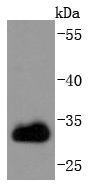
Figure 1. Western blot analysis of CDK1 using anti-CDK1 antibody (PB9533). Electrophoresis was performed on a 5-20% SDS-PAGE gel at 70V (Stacking gel) / 90V (Resolving gel) for 2-3 hours. Lane 1: Rat Thymus Tissue Lysate at 50ug, Lane 2: Rat Spleen Tissue Lysate at 50ug, Lane 3: MCF-7 Whole Cell Lysate at 40ug, Lane 4: HELA Whole Cell Lysate at 40ug, Lane 5: JURKAT Whole Cell Lysate at 40ug, Lane 6: NIH3T3 Whole Cell Lysate at 40ug. After electrophoresis, proteins were transferred to a nitrocellulose membrane at 150 mA for 50-90 minutes. Blocked the membrane with 5% non-fat milk/TBS for 1.5 hour at RT. The membrane was incubated with rabbit anti-CDK1 antigen affinity purified polyclonal antibody (Catalog # PB9533) at 0.5 microg/mL overnight at 4°C, then washed with TBS-0.1%Tween 3 times with 5 minutes each and probed with a goat anti-rabbit IgG-HRP secondary antibody at a dilution of 1:5000 for 1.5 hour at RT. The signal is developed using an Enhanced Chemiluminescent detection (ECL) kit (Catalog # EK1002)?with Tanon 5200 system. A specific band was detected for CDK1 at approximately 34 kDa. The expected band size for CDK1 is at 34 kDa.
. CDK1 was detected in a paraffin-embedded section of mouse testis tissue. Heat mediated antigen retrieval was performed in EDTA buffer (pH 8.0, epitope retrieval solution). The tissue section was blocked with 10% goat serum. The tissue section was then incubated with 1 microg/ml rabbit anti-CDK1 Antibody (PB9533) overnight at 4°C. Biotinylated goat anti-rabbit IgG was used as secondary antibody and incubated for 30 minutes at 37°C. The tissue section was developed using Strepavidin-Biotin-Complex (SABC) (Catalog # SA1022) with DAB as the chromogen.")
. CDK1 was detected in a paraffin-embedded section of rat testis tissue. Heat mediated antigen retrieval was performed in EDTA buffer (pH 8.0, epitope retrieval solution). The tissue section was blocked with 10% goat serum. The tissue section was then incubated with 1 microg/ml rabbit anti-CDK1 Antibody (PB9533) overnight at 4°C. Biotinylated goat anti-rabbit IgG was used as secondary antibody and incubated for 30 minutes at 37°C. The tissue section was developed using Strepavidin-Biotin-Complex (SABC) (Catalog # SA1022) with DAB as the chromogen.")
. CDK1 was detected in a paraffin-embedded section of human mammary cancer tissue. Heat mediated antigen retrieval was performed in EDTA buffer (pH 8.0, epitope retrieval solution). The tissue section was blocked with 10% goat serum. The tissue section was then incubated with 1 microg/ml rabbit anti-CDK1 Antibody (PB9533) overnight at 4°C. Biotinylated goat anti-rabbit IgG was used as secondary antibody and incubated for 30 minutes at 37°C. The tissue section was developed using Strepavidin-Biotin-Complex (SABC) (Catalog # SA1022) with DAB as the chromogen.")
. CDK1 was detected in immunocytochemical section of U20S cell. Enzyme antigen retrieval was performed using IHC enzyme antigen retrieval reagent (AR0022) for 15 mins. The cells were blocked with 10% goat serum. And then incubated with 2microg/mL rabbit anti-CDK1 Antibody (PB9533) overnight at 4°C. DyLight?488 Conjugated Goat Anti-Rabbit IgG (BA1127) was used as secondary antibody at 1:100 dilution and incubated for 30 minutes at 37°C. The section was counterstained with DAPI. Visualize using a fluorescence microscope and filter sets appropriate for the label used.")
. Overlay histogram showing U937 cells stained with PB9533 (Blue line).The cells were blocked with 10% normal goat serum. And then incubated with rabbit anti-CDK1 Antibody (PB9533,1microg/1x106 cells) for 30 min at 20°C. DyLight?488 conjugated goat anti-rabbit IgG (BA1127, 5-10microg/1x106 cells) was used as secondary antibody for 30 minutes at 20°C. Isotype control antibody (Green line) was rabbit IgG (1microg/1x106) used under the same conditions. Unlabelled sample (Red line) was also used as a control.")
Figure 1. Western blot analysis of CDK1 using anti-CDK1 antibody (PB9533). Electrophoresis was performed on a 5-20% SDS-PAGE gel at 70V (Stacking gel) / 90V (Resolving gel) for 2-3 hours. Lane 1: Rat Thymus Tissue Lysate at 50ug, Lane 2: Rat Spleen Tissue Lysate at 50ug, Lane 3: MCF-7 Whole Cell Lysate at 40ug, Lane 4: HELA Whole Cell Lysate at 40ug, Lane 5: JURKAT Whole Cell Lysate at 40ug, Lane 6: NIH3T3 Whole Cell Lysate at 40ug. After electrophoresis, proteins were transferred to a nitrocellulose membrane at 150 mA for 50-90 minutes. Blocked the membrane with 5% non-fat milk/TBS for 1.5 hour at RT. The membrane was incubated with rabbit anti-CDK1 antigen affinity purified polyclonal antibody (Catalog # PB9533) at 0.5 microg/mL overnight at 4°C, then washed with TBS-0.1%Tween 3 times with 5 minutes each and probed with a goat anti-rabbit IgG-HRP secondary antibody at a dilution of 1:5000 for 1.5 hour at RT. The signal is developed using an Enhanced Chemiluminescent detection (ECL) kit (Catalog # EK1002)?with Tanon 5200 system. A specific band was detected for CDK1 at approximately 34 kDa. The expected band size for CDK1 is at 34 kDa.
Anti-CDK1 Antibody Picoband(r)
PB9533-CARRIER-FREE
ApplicationsFlow Cytometry, ImmunoFluorescence, Western Blot, ImmunoCytoChemistry, ImmunoHistoChemistry, ImmunoHistoChemistry Frozen
Product group Antibodies
ReactivityHuman, Mouse, Rat
TargetCDK1
Overview
- SupplierBoster Bio
- Product NameAnti-CDK1 Antibody Picoband(r)
- Delivery Days Customer9
- Application Supplier NoteTested Species: In-house tested species with positive results. By Heat: Boiling the paraffin sections in 10mM citrate buffer, pH6.0, for 20mins is required for the staining of formalin/paraffin sections. Other applications have not been tested. Optimal dilutions should be determined by end users.
- ApplicationsFlow Cytometry, ImmunoFluorescence, Western Blot, ImmunoCytoChemistry, ImmunoHistoChemistry, ImmunoHistoChemistry Frozen
- CertificationResearch Use Only
- ClonalityPolyclonal
- Concentration500 ug/ml
- Gene ID983
- Target nameCDK1
- Target descriptioncyclin dependent kinase 1
- Target synonymsCDC2, CDC28A, P34CDC2, cyclin-dependent kinase 1, cell cycle controller CDC2, cell division control protein 2 homolog, cell division cycle 2, G1 to S and G2 to M, cell division protein kinase 1, p34 protein kinase
- HostRabbit
- IsotypeIgG
- Protein IDP06493
- Protein NameCyclin-dependent kinase 1
- Scientific DescriptionBoster Bio Anti-CDK1 Antibody Picoband® catalog # PB9533. Tested in Flow Cytometry, IF, IHC, IHC-F, ICC, WB applications. This antibody reacts with Human, Mouse, Rat. The brand Picoband indicates this is a premium antibody that guarantees superior quality, high affinity, and strong signals with minimal background in Western blot applications. Only our best-performing antibodies are designated as Picoband, ensuring unmatched performance.
- ReactivityHuman, Mouse, Rat
- Storage Instruction-20°C,2°C to 8°C
- UNSPSC12352203
Related products
Product group Antibodies
CDK1 AntibodyCSB-PA001495
ApplicationsWestern Blot, ELISA
ReactivityHuman, Mouse, Rat
TargetCDK1
- SizePrice
Product group Antibodies
Anti-CDC2 AntibodyA96412
ApplicationsImmunoFluorescence, Western Blot, ELISA
ReactivityHuman, Mouse, Rat
- SizePrice
Product group Antibodies

CDK1 / CDC2 AntibodyLS-C765432
ApplicationsWestern Blot
ReactivityHuman
TargetCDK1
- SizePrice

Product group Antibodies
Anti-CDK1 AntibodyHPA003387
ApplicationsWestern Blot, ImmunoCytoChemistry, ImmunoHistoChemistry
ReactivityHuman, Mouse, Rat
TargetCDK1
- SizePrice
Product group Antibodies
ApplicationsImmunoPrecipitation, Western Blot, ImmunoCytoChemistry, ImmunoHistoChemistry
ReactivityMouse, Rat
TargetCDK1
- SizePrice
Product group Antibodies
CDK1 Recombinant AntibodyBSM-52026R
ApplicationsFlow Cytometry, ImmunoFluorescence, Western Blot, ImmunoCytoChemistry, ImmunoHistoChemistry, ImmunoHistoChemistry Frozen, ImmunoHistoChemistry Paraffin
ReactivityHuman, Mouse, Rat
TargetCDK1
- SizePrice
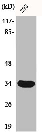
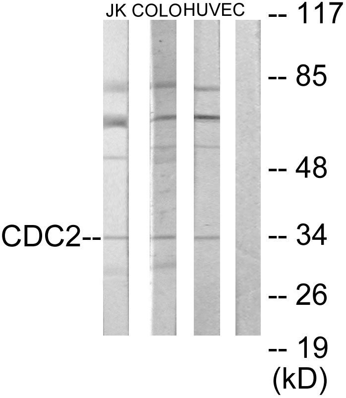
![Various whole cell extracts (30 μg) were separated by 10% SDS-PAGE, and the membrane was blotted with CDC2 antibody [N1], N-term (GTX108120) diluted at 1:1000. The HRP-conjugated anti-rabbit IgG antibody (GTX213110-01) was used to detect the primary antibody.](https://www.genetex.com/upload/website/prouct_img/normal/GTX108120/GTX108120_39778_20210528_WB_w_23060120_585.webp)
Product group Antibodies
CDC2 antibody [N1], N-termGTX108120
ApplicationsImmunoFluorescence, ImmunoPrecipitation, Western Blot, ImmunoCytoChemistry, ImmunoHistoChemistry, ImmunoHistoChemistry Paraffin
ReactivityHuman, Mouse, Rat
TargetCDK1
- SizePrice