


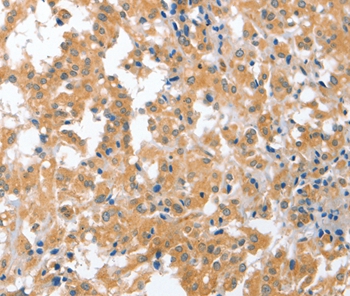
Immunohistochemical staining of human testis shows strong cytoplasmic and nuclear positivity in cells in seminiferous ducts.
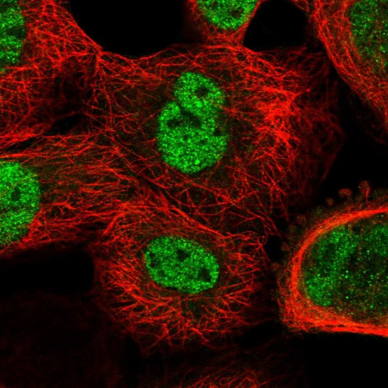
 shows similar pattern to independent antibody HPA023870 (B).")
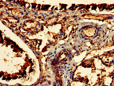
Immunohistochemical staining of human testis shows strong cytoplasmic and nuclear positivity in cells in seminiferous ducts.
Anti-CDR2 Antibody
HPA018151
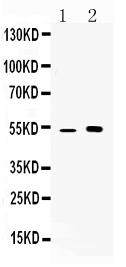
ApplicationsWestern Blot, ImmunoHistoChemistry
Product group Antibodies
ReactivityHuman
TargetCDR2
Overview
- SupplierAtlas Antibodies
- Product NameAnti-CDR2 Antibody
- Delivery Days Customer4
- ApplicationsWestern Blot, ImmunoHistoChemistry
- CertificationResearch Use Only
- ClonalityPolyclonal
- ConjugateUnconjugated
- Gene ID1039
- Target nameCDR2
- Target descriptioncerebellar degeneration related protein 2
- Target synonymsCDR62, Yo, cerebellar degeneration-related protein 2, Yo paraneoplastic antigen, cerebellar degeneration-related protein 2, 62kDa, major Yo paraneoplastic antigen, paraneoplastic cerebellar degeneration-associated antigen
- HostRabbit
- IsotypeIgG
- Protein IDQ01850
- Protein NameCerebellar degeneration-related protein 2
- Scientific DescriptionRecombinant Protein Epitope Signature Tag (PrEST) antigen sequence
- ReactivityHuman
- Storage Instruction-20°C,2°C to 8°C
- UNSPSC41116161
Datasheet
MSDS
Related products
Product group Antibodies
Anti-CDR2 AntibodyA46783
ApplicationsImmunoHistoChemistry
ReactivityHuman
- SizePrice
Product group Antibodies
Anti-CDR2 Antibody101-10269
ApplicationsImmunoFluorescence, Western Blot, ELISA
TargetCDR2
- SizePrice
Product group Antibodies
Anti-CDR2 Antibody Picoband(r)A02973-1-CARRIER-FREE
ApplicationsWestern Blot
ReactivityHuman, Mouse
TargetCDR2
- SizePrice
Product group Antibodies
Cdr2 Polyclonal AntibodyCAC08306
ApplicationsWestern Blot, ELISA, ImmunoHistoChemistry
ReactivityMouse
TargetCDR2
- SizePrice
Product group Antibodies
CDR2 AntibodyCSB-PA005103LA01HU
ApplicationsWestern Blot, ELISA, ImmunoHistoChemistry
ReactivityHuman, Mouse
TargetCDR2
- SizePrice
Product group Antibodies

Yo / CDR2 AntibodyLS-C401514
ApplicationsELISA, ImmunoHistoChemistry
ReactivityHuman, Mouse
TargetCDR2
- SizePrice
Product group Antibodies
Anti-CDR2 AntibodyHPA023870
ApplicationsWestern Blot, ImmunoCytoChemistry
ReactivityHuman
TargetCDR2
- SizePrice