


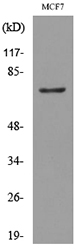
Figure 1. Western blot analysis of CEA using anti-CEA antibody (RP1018). Electrophoresis was performed on a 5-20% SDS-PAGE gel at 70V (Stacking gel) / 90V (Resolving gel) for 2-3 hours. The sample well of each lane was loaded with 50ug of sample under reducing conditions. Lane 1: human SW620 whole cell lysates, Lane 2: human Caco-2 whole cell lysates, Lane 3: mouse small intestine tissue lysates, Lane 4: mouse stomach tissue lysates, Lane 5: mouse lung tissue lysates, Lane 6: mouse liver tissue lysates, Lane 7: mouse NIH3T3 whole cell lysates, Lane 8: mouse HEPA1-6 whole cell lysates, Lane 9: mouse SP20 whole cell lysates, Lane 10: rat RH35 whole cell lysates. After Electrophoresis, proteins were transferred to a Nitrocellulose membrane at 150mA for 50-90 minutes. Blocked the membrane with 5% Non-fat Milk/ TBS for 1.5 hour at RT. The membrane was incubated with rabbit anti-CEA antigen affinity purified polyclonal antibody (Catalog # RP1018) at 0.5 microg/mL overnight at 4°C, then washed with TBS-0.1%Tween 3 times with 5 minutes each and probed with a goat anti-rabbit IgG-HRP secondary antibody at a dilution of 1:10000 for 1.5 hour at RT. The signal is developed using an Enhanced Chemiluminescent detection (ECL) kit (Catalog # EK1002) with Tanon 5200 system. A specific band was detected for CEA at approximately 120-200KD. The expected band size for CEA is at 77KD.
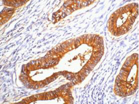
. CEA was detected in a paraffin-embedded section of human colon cancer tissue. Heat mediated antigen retrieval was performed in EDTA buffer (pH 8.0, epitope retrieval solution). The tissue section was blocked with 10% goat serum. The tissue section was then incubated with 1 microg/ml rabbit anti-CEA Antibody (RP1018) overnight at 4°C. Peroxidase Conjugated Goat Anti-rabbit IgG was used as secondary antibody and incubated for 30 minutes at 37°C. The tissue section was developed using HRP Conjugated Rabbit IgG Super Vision Assay Kit (Catalog # SV0002) with DAB as the chromogen.")
Figure 1. Western blot analysis of CEA using anti-CEA antibody (RP1018). Electrophoresis was performed on a 5-20% SDS-PAGE gel at 70V (Stacking gel) / 90V (Resolving gel) for 2-3 hours. The sample well of each lane was loaded with 50ug of sample under reducing conditions. Lane 1: human SW620 whole cell lysates, Lane 2: human Caco-2 whole cell lysates, Lane 3: mouse small intestine tissue lysates, Lane 4: mouse stomach tissue lysates, Lane 5: mouse lung tissue lysates, Lane 6: mouse liver tissue lysates, Lane 7: mouse NIH3T3 whole cell lysates, Lane 8: mouse HEPA1-6 whole cell lysates, Lane 9: mouse SP20 whole cell lysates, Lane 10: rat RH35 whole cell lysates. After Electrophoresis, proteins were transferred to a Nitrocellulose membrane at 150mA for 50-90 minutes. Blocked the membrane with 5% Non-fat Milk/ TBS for 1.5 hour at RT. The membrane was incubated with rabbit anti-CEA antigen affinity purified polyclonal antibody (Catalog # RP1018) at 0.5 microg/mL overnight at 4°C, then washed with TBS-0.1%Tween 3 times with 5 minutes each and probed with a goat anti-rabbit IgG-HRP secondary antibody at a dilution of 1:10000 for 1.5 hour at RT. The signal is developed using an Enhanced Chemiluminescent detection (ECL) kit (Catalog # EK1002) with Tanon 5200 system. A specific band was detected for CEA at approximately 120-200KD. The expected band size for CEA is at 77KD.
Anti-CEACAM5 Antibody Picoband(r)
RP1018
ApplicationsWestern Blot, ELISA, ImmunoHistoChemistry
Product group Antibodies
TargetCEACAM5
Overview
- SupplierBoster Bio
- Product NameAnti-CEA Antibody
- Delivery Days Customer9
- Application Supplier NoteBy Heat: Boiling the paraffin sections in 10mM citrate buffer, pH6.0, for 20mins is required for the staining of formalin/paraffin sections. Other applications have not been tested. Optimal dilutions should be determined by end users.
- ApplicationsWestern Blot, ELISA, ImmunoHistoChemistry
- Applications SupplierELI, IHP, WB, IHC
- CertificationResearch Use Only
- ClonalityPolyclonal
- Concentration500 ug/ml
- Gene ID1048
- Target nameCEACAM5
- Target descriptionCEA cell adhesion molecule 5
- Target synonymscarcinoembryonic antigen related cell adhesion molecule 5; carcinoembryonic antigen-related cell adhesion molecule 5; CD66e; CEA; meconium antigen 100
- HostRabbit
- IsotypeIgG
- Protein IDP06731
- Protein NameCarcinoembryonic antigen-related cell adhesion molecule 5
- Scientific DescriptionBoster Bio Anti-CEACAM5 Antibody catalog # RP1018. Tested in ELISA, IHC, WB applications. This antibody reacts with Human. The brand Picoband indicates this is a premium antibody that guarantees superior quality, high affinity, and strong signals with minimal background in Western blot applications. Only our best-performing antibodies are designated as Picoband, ensuring unmatched performance.
- Reactivity SupplierHuman
- Storage Instruction-20°C,2°C to 8°C
- UNSPSC12352203
References
- Band-Edge Effect-Induced Electrochemiluminescence Signal Amplification Based on Inverse Opal Photonic Crystals for Ultrasensitive Detection of Carcinoembryonic Antigen.Read more
- An ultrasensitive homogeneous aptasensor for carcinoembryonic antigen based on upconversion fluorescence resonance energy transfer.Read more
Datasheet
MSDS
Related products
Product group Antibodies
Anti-CEACAM5 Antibody144-60045
ApplicationsWestern Blot
TargetCEACAM5
- SizePrice
Product group Antibodies
ApplicationsWestern Blot
TargetCEACAM5
- SizePrice
Product group Antibodies

CEACAM5 / CD66e Antibody (clone 1A2)LS-C765377
ApplicationsImmunoFluorescence, ImmunoHistoChemistry, ImmunoHistoChemistry Paraffin
TargetCEACAM5
- SizePrice

Product group Antibodies
CEACAM5 Monoclonal AntibodyCSB-MA000218
ApplicationsELISA, ImmunoHistoChemistry
ReactivityHuman
TargetCEACAM5
- SizePrice
Product group Antibodies
ApplicationsImmunoPrecipitation, Western Blot, ImmunoCytoChemistry, ImmunoHistoChemistry
TargetCEACAM5
- SizePrice
Product group Antibodies
References
CEA(B5) Monoclonal AntibodyBSM-1624M
ApplicationsImmunoFluorescence, Western Blot, ELISA, ImmunoHistoChemistry, ImmunoHistoChemistry Paraffin
TargetCEACAM5
- SizePrice