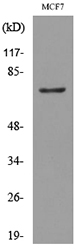
Western blot analysis of CEA(CD66e) expression in Human colon cancer lysate.

 Antibody.")

 Antibody.")
Western blot analysis of CEA(CD66e) expression in Human colon cancer lysate.
Anti-CEA (CD66e) CEACAM5 Rabbit Monoclonal Antibody
M00356-1
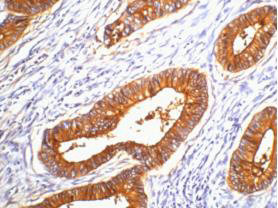
ApplicationsImmunoFluorescence, Western Blot, ImmunoCytoChemistry, ImmunoHistoChemistry
Product group Antibodies
TargetCEACAM5
Overview
- SupplierBoster Bio
- Product NameAnti-CEA (CD66e) Rabbit Monoclonal Antibody
- Delivery Days Customer9
- ApplicationsImmunoFluorescence, Western Blot, ImmunoCytoChemistry, ImmunoHistoChemistry
- CertificationResearch Use Only
- ClonalityMonoclonal
- Clone IDAAOA-3
- Gene ID1048
- Target nameCEACAM5
- Target descriptionCEA cell adhesion molecule 5
- Target synonymsCD66e, CEA, cell adhesion molecule CEACAM5, carcinoembryonic antigen related cell adhesion molecule 5, meconium antigen 100
- HostRabbit
- IsotypeIgG
- Protein IDP06731
- Protein NameCarcinoembryonic antigen-related cell adhesion molecule 5
- Scientific DescriptionBoster Bio Anti-CEA (CD66e) CEACAM5 Rabbit Monoclonal Antibody catalog # M00356-1. Tested in WB, IHC, ICC/IF applications. This antibody reacts with Human.
- Storage Instruction-20°C
- UNSPSC12352203
Datasheet
MSDS
Related products
Product group Antibodies
ApplicationsWestern Blot
TargetCEACAM5
- SizePrice
Product group Antibodies
CEACAM5 Monoclonal AntibodyCSB-MA000218
ApplicationsELISA, ImmunoHistoChemistry
ReactivityHuman
TargetCEACAM5
- SizePrice
Product group Antibodies
References
CD66e antibodyGTX100903
ApplicationsImmunoFluorescence, Western Blot, ImmunoCytoChemistry
TargetCEACAM5
- SizePrice

Product group Antibodies
ApplicationsImmunoPrecipitation, Western Blot, ImmunoCytoChemistry, ImmunoHistoChemistry
TargetCEACAM5
- SizePrice

Product group Antibodies
Anti-CEA/CEACAM5 Antibody Picoband(r)A00356-2-CARRIER-FREE
ApplicationsFlow Cytometry, Western Blot, ELISA
TargetCEACAM5
- SizePrice
Product group Antibodies
Anti-CEACAM5 Antibody144-60045
ApplicationsWestern Blot
TargetCEACAM5
- SizePrice