

Anti-CEA [T84.66]
Ab02833-10.0
ApplicationsFlow Cytometry, ELISA, ImmunoHistoChemistry, RadioImmunoAssay
Product group Antibodies
TargetCEACAM5
Overview
- SupplierAbsolute Antibody
- Product NameAnti-CEA [T84.66]
- Delivery Days Customer7
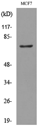
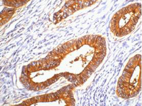
- Application Supplier NoteThe binding affinity of the original mouse antibody to carcinoembryonic antigen was determined using a competitive radioimmunoassay (PMID: 6187848). 111In (Indacea) labelled version of this antibody was used to image tumors in patients with colorectal cancer (PMID: 3779658). An engineered antibody fragment (VL-VH-CH3, or minibody) generated using this antibody having bivalent binding to carcinoembryonic antigen showed high level targeting of xenografts (PMID: 8674062). A radiolabeled chimeric version of this antibody was used to target breast cancer cells expressing high levels of CEA and it was found that chimeric 86.66 resulted in good tumor localization as well as significant tumor growth inhibition. This antibody was also used to detect CEA expression in Human breast carcinoma cell line, MCF 7, using flow cytometry and immunohistochemistry (PMID: 21597719). The characterization of the chimeric version of this antibody generated by transient expression in tobacco leaves was done using ELISA (PMID: 10500141). A phase 1 clinical radioimmunotherapy trial studying 22 patients with metastatic CEA-producing malignancies involving 90Yttrium-labeled chimeric T86.44 revealed that the antibody was well tolerated, with reversible thrombocytopenia and leukopenia being dose limiting (PMID: 11051230). This antibody was used in the immunohistochemical staining of paraffin sections of primary tumours from 252 consecutive patients with breast carcinomas (PMID: 9635845).
- ApplicationsFlow Cytometry, ELISA, ImmunoHistoChemistry, RadioImmunoAssay
- Applications SupplierELISA; RIA; FC; IHC; Immunoscintigraphy; immunotherapy
- CertificationResearch Use Only
- ClonalityMonoclonal
- Clone IDT84.66
- Gene ID1048
- Target nameCEACAM5
- Target descriptionCEA cell adhesion molecule 5
- Target synonymscarcinoembryonic antigen related cell adhesion molecule 5; carcinoembryonic antigen-related cell adhesion molecule 5; CD66e; CEA; meconium antigen 100
- HostHuman
- IsotypeIgG1
- Protein IDP06731
- Protein NameCarcinoembryonic antigen-related cell adhesion molecule 5
- Scientific DescriptionThis chimeric human antibody was made using the variable domain sequences of the original Mouse IgG1 format for improved compatibility with existing reagents assays and techniques.
- Reactivity SupplierHuman
- Reactivity Supplier NoteThe original antibody was generated by immunizing female BALB/c mice subcutaneously with soluble CEA or with the HC84S cell line.
- Storage Instruction-20°C,2°C to 8°C
- UNSPSC12352203
Related products
Product group Antibodies
Anti-CEACAM5 Antibody144-60045
ApplicationsWestern Blot
TargetCEACAM5
- SizePrice
Product group Antibodies
ApplicationsWestern Blot
TargetCEACAM5
- SizePrice
Product group Antibodies

CEACAM5 / CD66e Antibody (clone 1A2)LS-C765377
ApplicationsImmunoFluorescence, ImmunoHistoChemistry, ImmunoHistoChemistry Paraffin
TargetCEACAM5
- SizePrice

Product group Antibodies
CEACAM5 Monoclonal AntibodyCSB-MA000218
ApplicationsELISA, ImmunoHistoChemistry
ReactivityHuman
TargetCEACAM5
- SizePrice
Product group Antibodies
ApplicationsImmunoPrecipitation, Western Blot, ImmunoCytoChemistry, ImmunoHistoChemistry
TargetCEACAM5
- SizePrice
Product group Antibodies
References
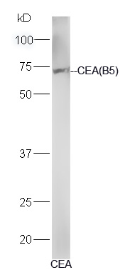
CEA(B5) Monoclonal AntibodyBSM-1624M
ApplicationsImmunoFluorescence, Western Blot, ELISA, ImmunoHistoChemistry, ImmunoHistoChemistry Paraffin
TargetCEACAM5
- SizePrice