

Anti-CEACAM5 [NbCEA5], Human IgG1-Fc Fusion,
AB04388-10.159-BT
ApplicationsFlow Cytometry, ELISA, Other Application
Product group Antibodies
ReactivityHuman
TargetCEACAM5
Product AB04388-10.159-BT is not available
Product not available
There may be an alternative product available, please contact our technical support team.
Overview
- SupplierAbsolute Antibody
- Product NameAnti-CEACAM5 [NbCEA5], Human IgG1-Fc Fusion,
- Delivery Days Customer9
- Application Supplier NoteThe binding of the original version of this antibody (VHH) to CEACAM5 was characterized in an SPR (KD 0.41 nM), ELISA (EC50 of 0.026 nM), and FACS (EC50 of 1.0 nM). In its bi-specific construction with Ab04389 (Anti-EGFR [7D12]), it served as an anchor (US20220242958).
- ApplicationsFlow Cytometry, ELISA, Other Application
- CertificationResearch Use Only
- ClonalityMonoclonal
- Clone IDNbCEA5
- Gene ID1048
- Target nameCEACAM5
- Target descriptionCEA cell adhesion molecule 5
- Target synonymsCD66e, CEA, cell adhesion molecule CEACAM5, carcinoembryonic antigen related cell adhesion molecule 5, meconium antigen 100
- HostHuman
- IsotypeIgG1
- Protein IDP06731
- Protein NameCell adhesion molecule CEACAM5
- Scientific DescriptionThis chimeric human antibody was made using the variable domain sequences of the original VHH format, for improved compatibility with existing reagents, assays and techniques.
- ReactivityHuman
- Storage Instruction-20°C,2°C to 8°C
- UNSPSC12352203
Related products
Product group Antibodies

Anti-CEA [EB-011]Ab01002-1.1
ApplicationsELISA
ReactivityHuman
TargetCEACAM5
- SizePrice
Product group Antibodies
Anti-CEACAM5 Antibody144-60045
ApplicationsWestern Blot
ReactivityHuman, Mouse
TargetCEACAM5
- SizePrice
Product group Antibodies
ApplicationsWestern Blot
ReactivityMouse
TargetCEACAM5
- SizePrice
Product group Antibodies
References
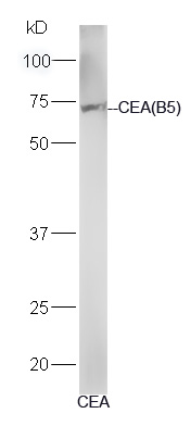
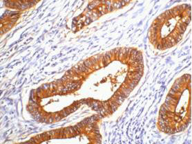
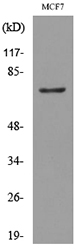
CEA(B5) Monoclonal AntibodyBSM-1624M
ApplicationsImmunoFluorescence, Western Blot, ELISA, ImmunoHistoChemistry, ImmunoHistoChemistry Paraffin
ReactivityHuman
TargetCEACAM5
- SizePrice
Product group Antibodies
CEACAM5 Monoclonal AntibodyCSB-MA000218
ApplicationsELISA, ImmunoHistoChemistry
ReactivityHuman
TargetCEACAM5
- SizePrice
Product group Antibodies

CEACAM5 / CD66e Antibody (clone 1A2)LS-C765377
ApplicationsImmunoFluorescence, ImmunoHistoChemistry, ImmunoHistoChemistry Paraffin
ReactivityHuman
TargetCEACAM5
- SizePrice
Product group Antibodies
Anti-CEACAM5 AntibodyA100175
ApplicationsWestern Blot, ELISA
ReactivityHuman
- SizePrice