


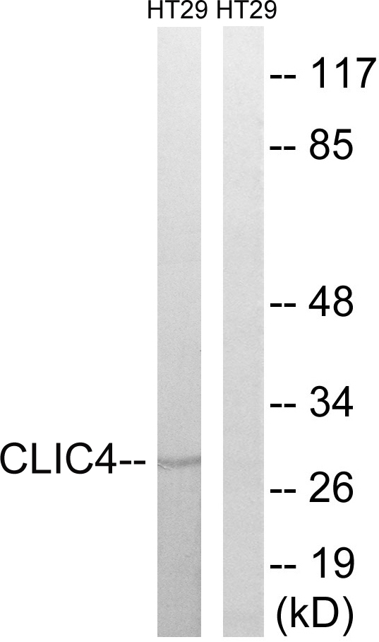
Figure 1. Western blot analysis of CLIC4 using anti-CLIC4 antibody (M03210). Electrophoresis was performed on a 5-20% SDS-PAGE gel at 70V (Stacking gel) / 90V (Resolving gel) for 2-3 hours. The sample well of each lane was loaded with 30 ug of sample under reducing conditions. Lane 1: human 293T whole cell lysates, Lane 2: human U251 whole cell lysates, Lane 3: human A431 whole cell lysates, Lane 4: human U20S whole cell lysates, Lane 5: rat kidney tissue lysates, Lane 6: rat brain tissue lysates, Lane 7: mouse kidney tissue lysates, Lane 8: mouse brain tissue lysates. After electrophoresis, proteins were transferred to a nitrocellulose membrane at 150 mA for 50-90 minutes. Blocked the membrane with 5% non-fat milk/TBS for 1.5 hour at RT. The membrane was incubated with rabbit anti-CLIC4 antigen affinity purified monoclonal antibody (Catalog # M03210) at 1:500 overnight at 4°C, then washed with TBS-0.1%Tween 3 times with 5 minutes each and probed with a goat anti-rabbit IgG-HRP secondary antibody at a dilution of 1:500 for 1.5 hour at RT. The signal is developed using an Enhanced Chemiluminescent detection (ECL) kit (Catalog # EK1002) with Tanon 5200 system. A specific band was detected for CLIC4 at approximately 29 kDa. The expected band size for CLIC4 is at 29 kDa.
Figure 1. Western blot analysis of CLIC4 using anti-CLIC4 antibody (M03210). Electrophoresis was performed on a 5-20% SDS-PAGE gel at 70V (Stacking gel) / 90V (Resolving gel) for 2-3 hours. The sample well of each lane was loaded with 30 ug of sample under reducing conditions. Lane 1: human 293T whole cell lysates, Lane 2: human U251 whole cell lysates, Lane 3: human A431 whole cell lysates, Lane 4: human U20S whole cell lysates, Lane 5: rat kidney tissue lysates, Lane 6: rat brain tissue lysates, Lane 7: mouse kidney tissue lysates, Lane 8: mouse brain tissue lysates. After electrophoresis, proteins were transferred to a nitrocellulose membrane at 150 mA for 50-90 minutes. Blocked the membrane with 5% non-fat milk/TBS for 1.5 hour at RT. The membrane was incubated with rabbit anti-CLIC4 antigen affinity purified monoclonal antibody (Catalog # M03210) at 1:500 overnight at 4°C, then washed with TBS-0.1%Tween 3 times with 5 minutes each and probed with a goat anti-rabbit IgG-HRP secondary antibody at a dilution of 1:500 for 1.5 hour at RT. The signal is developed using an Enhanced Chemiluminescent detection (ECL) kit (Catalog # EK1002) with Tanon 5200 system. A specific band was detected for CLIC4 at approximately 29 kDa. The expected band size for CLIC4 is at 29 kDa.
Anti-CLIC4 Rabbit Monoclonal Antibody
M03210
ApplicationsFlow Cytometry, ImmunoFluorescence, ImmunoPrecipitation, Western Blot, ImmunoCytoChemistry, ImmunoHistoChemistry
Product group Antibodies
ReactivityHuman, Mouse, Rat
TargetCLIC4
Overview
- SupplierBoster Bio
- Product NameAnti-CLIC4 Rabbit Monoclonal Antibody
- Delivery Days Customer9
- ApplicationsFlow Cytometry, ImmunoFluorescence, ImmunoPrecipitation, Western Blot, ImmunoCytoChemistry, ImmunoHistoChemistry
- CertificationResearch Use Only
- ClonalityMonoclonal
- Clone ID30C24
- Gene ID25932
- Target nameCLIC4
- Target descriptionchloride intracellular channel 4
- Target synonymsCLIC4L, H1, MTCLIC, huH1, p64H1, chloride intracellular channel protein 4, chloride intracellular channel 4 like, epididymis secretory sperm binding protein, glutaredoxin-like oxidoreductase CLIC4, intracellular chloride ion channel protein p64H1
- HostRabbit
- IsotypeIgG
- Protein IDQ9Y696
- Protein NameChloride intracellular channel protein 4
- Scientific DescriptionBoster Bio Anti-CLIC4 Rabbit Monoclonal Antibody catalog # M03210. Tested in WB, IHC, ICC/IF, IP, Flow Cytometry applications. This antibody reacts with Human, Mouse, Rat.
- ReactivityHuman, Mouse, Rat
- Storage Instruction-20°C
- UNSPSC12352203
Related products
Product group Antibodies
Anti-CLIC4 Antibody144-07088
ApplicationsImmunoFluorescence, Western Blot, ImmunoHistoChemistry
ReactivityHuman, Mouse, Rat
TargetCLIC4
- SizePrice
Product group Antibodies
Anti-CLIC4 AntibodyA95906
ApplicationsWestern Blot, ELISA, ImmunoHistoChemistry
ReactivityHuman, Mouse, Rat
- SizePrice
Product group Antibodies
ApplicationsImmunoPrecipitation, Western Blot, ImmunoCytoChemistry, ImmunoHistoChemistry
ReactivityMouse, Rat
TargetCLIC4
- SizePrice
Product group Antibodies
CLIC4 AntibodyCSB-PA001691
ApplicationsWestern Blot, ELISA, ImmunoHistoChemistry
ReactivityHuman, Mouse, Rat
TargetCLIC4
- SizePrice
Product group Antibodies
CLIC4 antibodyGTX104888
ApplicationsImmunoFluorescence, Western Blot, ImmunoCytoChemistry, ImmunoHistoChemistry, ImmunoHistoChemistry Paraffin
ReactivityHuman, Zebra Fish
TargetCLIC4
- SizePrice

Product group Antibodies
Anti-CLIC4 AntibodyHPA008019
ApplicationsWestern Blot, ImmunoHistoChemistry
ReactivityHuman
TargetCLIC4
- SizePrice
Product group Antibodies

Goat anti-CLIC4EB07738
ApplicationsImmunoFluorescence, Western Blot, ELISA, ImmunoHistoChemistry
ReactivityCanine, Human, Mouse, Rat
TargetCLIC4
- SizePrice
Product group Antibodies

CLIC4 AntibodyLS-C832629
ApplicationsWestern Blot, ELISA
ReactivityHuman, Mouse, Rat
TargetCLIC4
- SizePrice
Product group Antibodies
ApplicationsFlow Cytometry, Western Blot, ImmunoCytoChemistry
ReactivityHuman, Mouse, Rat
TargetCLIC4
- SizePrice