


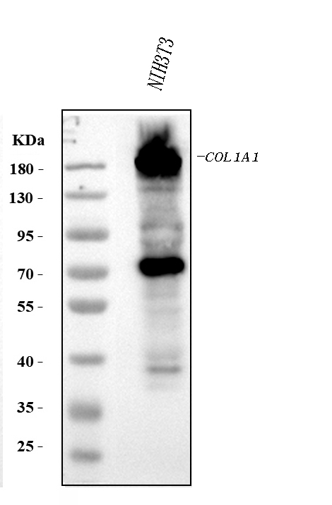
Figure 1. Western blot analysis of Collagen I/COL1A1 using anti-Collagen I/COL1A1 antibody (PA2140-2). Electrophoresis was performed on a 5-20% SDS-PAGE gel at 70V (Stacking gel) / 90V (Resolving gel) for 2-3 hours. The sample well of each lane was loaded with 30 ug of sample under reducing conditions. Lane 1: human placenta tissue lysates, Lane 2: rat skin tissue lysates, Lane 3: rat lung tissue lysates, Lane 4: mouse skin tissue lysates, Lane 5: mouse NIH/3T3 whole cell lysates. After electrophoresis, proteins were transferred to a nitrocellulose membrane at 150 mA for 50-90 minutes. Blocked the membrane with 5% non-fat milk/TBS for 1.5 hour at RT. The membrane was incubated with rabbit anti-Collagen I/COL1A1 antigen affinity purified polyclonal antibody (Catalog # PA2140-2) at 0.5 microg/mL overnight at 4°C, then washed with TBS-0.1%Tween 3 times with 5 minutes each and probed with a goat anti-rabbit IgG-HRP secondary antibody at a dilution of 1:5000 for 1.5 hour at RT. The signal is developed using an Enhanced Chemiluminescent detection (ECL) kit (Catalog # EK1002) with Tanon 5200 system. A specific band was detected for Collagen I/COL1A1 at approximately 130 kDa, 220 kDa. The expected band size for Collagen I/COL1A1 is at 138 kDa.
. Collagen I/COL1A1 was detected in a paraffin-embedded section of human liver cancer tissue. Heat mediated antigen retrieval was performed in EDTA buffer (pH 8.0, epitope retrieval solution). The tissue section was blocked with 10% goat serum. The tissue section was then incubated with 2 microg/ml rabbit anti-Collagen I/COL1A1 Antibody (PA2140-2) overnight at 4°C. Peroxidase Conjugated Goat Anti-rabbit IgG was used as secondary antibody and incubated for 30 minutes at 37°C. The tissue section was developed using HRP Conjugated Rabbit IgG Super Vision Assay Kit (Catalog # SV0002) with DAB as the chromogen.")
. Collagen I/COL1A1 was detected in a paraffin-embedded section of human lung adenocarcinoma cancer tissue. Heat mediated antigen retrieval was performed in EDTA buffer (pH 8.0, epitope retrieval solution). The tissue section was blocked with 10% goat serum. The tissue section was then incubated with 2 microg/ml rabbit anti-Collagen I/COL1A1 Antibody (PA2140-2) overnight at 4°C. Peroxidase Conjugated Goat Anti-rabbit IgG was used as secondary antibody and incubated for 30 minutes at 37°C. The tissue section was developed using HRP Conjugated Rabbit IgG Super Vision Assay Kit (Catalog # SV0002) with DAB as the chromogen.")
. Collagen I/COL1A1 was detected in a paraffin-embedded section of human renal cell carcinoma tissue. Heat mediated antigen retrieval was performed in EDTA buffer (pH 8.0, epitope retrieval solution). The tissue section was blocked with 10% goat serum. The tissue section was then incubated with 2 microg/ml rabbit anti-Collagen I/COL1A1 Antibody (PA2140-2) overnight at 4°C. Peroxidase Conjugated Goat Anti-rabbit IgG was used as secondary antibody and incubated for 30 minutes at 37°C. The tissue section was developed using HRP Conjugated Rabbit IgG Super Vision Assay Kit (Catalog # SV0002) with DAB as the chromogen.")
. Collagen I/COL1A1 was detected in a paraffin-embedded section of human bladder urothelial carcinoma tissue. Heat mediated antigen retrieval was performed in EDTA buffer (pH 8.0, epitope retrieval solution). The tissue section was blocked with 10% goat serum. The tissue section was then incubated with 2 microg/ml rabbit anti-Collagen I/COL1A1 Antibody (PA2140-2) overnight at 4°C. Peroxidase Conjugated Goat Anti-rabbit IgG was used as secondary antibody and incubated for 30 minutes at 37°C. The tissue section was developed using HRP Conjugated Rabbit IgG Super Vision Assay Kit (Catalog # SV0002) with DAB as the chromogen.")
. Collagen I/COL1A1 was detected in a paraffin-embedded section of human placenta tissue. Heat mediated antigen retrieval was performed in EDTA buffer (pH 8.0, epitope retrieval solution). The tissue section was blocked with 10% goat serum. The tissue section was then incubated with 2 microg/ml rabbit anti-Collagen I/COL1A1 Antibody (PA2140-2) overnight at 4°C. Peroxidase Conjugated Goat Anti-rabbit IgG was used as secondary antibody and incubated for 30 minutes at 37°C. The tissue section was developed using HRP Conjugated Rabbit IgG Super Vision Assay Kit (Catalog # SV0002) with DAB as the chromogen.")
. Collagen I/COL1A1 was detected in a paraffin-embedded section of mouse lung tissue. Heat mediated antigen retrieval was performed in EDTA buffer (pH 8.0, epitope retrieval solution). The tissue section was blocked with 10% goat serum. The tissue section was then incubated with 2 microg/ml rabbit anti-Collagen I/COL1A1 Antibody (PA2140-2) overnight at 4°C. Peroxidase Conjugated Goat Anti-rabbit IgG was used as secondary antibody and incubated for 30 minutes at 37°C. The tissue section was developed using HRP Conjugated Rabbit IgG Super Vision Assay Kit (Catalog # SV0002) with DAB as the chromogen.")
. Collagen I/COL1A1 was detected in a paraffin-embedded section of rat lung tissue. Heat mediated antigen retrieval was performed in EDTA buffer (pH 8.0, epitope retrieval solution). The tissue section was blocked with 10% goat serum. The tissue section was then incubated with 2 microg/ml rabbit anti-Collagen I/COL1A1 Antibody (PA2140-2) overnight at 4°C. Peroxidase Conjugated Goat Anti-rabbit IgG was used as secondary antibody and incubated for 30 minutes at 37°C. The tissue section was developed using HRP Conjugated Rabbit IgG Super Vision Assay Kit (Catalog # SV0002) with DAB as the chromogen.")
. Collagen I/COL1A1 was detected in a paraffin-embedded section of human placenta tissue. Heat mediated antigen retrieval was performed in EDTA buffer (pH 8.0, epitope retrieval solution). The tissue section was blocked with 10% goat serum. The tissue section was then incubated with 5 microg/mL rabbit anti-Collagen I/COL1A1 Antibody (PA2140-2) overnight at 4°C. Cy3 Conjugated Goat Anti-Rabbit IgG (BA1032) was used as secondary antibody at 1:500 dilution and incubated for 30 minutes at 37°C. The section was counterstained with DAPI. Visualize using a fluorescence microscope and filter sets appropriate for the label used.")
. Collagen I/COL1A1 was detected in a paraffin-embedded section of mouse lung tissue. Heat mediated antigen retrieval was performed in EDTA buffer (pH 8.0, epitope retrieval solution). The tissue section was blocked with 10% goat serum. The tissue section was then incubated with 5 microg/mL rabbit anti-Collagen I/COL1A1 Antibody (PA2140-2) overnight at 4°C. Cy3 Conjugated Goat Anti-Rabbit IgG (BA1032) was used as secondary antibody at 1:500 dilution and incubated for 30 minutes at 37°C. The section was counterstained with DAPI. Visualize using a fluorescence microscope and filter sets appropriate for the label used.")
Figure 1. Western blot analysis of Collagen I/COL1A1 using anti-Collagen I/COL1A1 antibody (PA2140-2). Electrophoresis was performed on a 5-20% SDS-PAGE gel at 70V (Stacking gel) / 90V (Resolving gel) for 2-3 hours. The sample well of each lane was loaded with 30 ug of sample under reducing conditions. Lane 1: human placenta tissue lysates, Lane 2: rat skin tissue lysates, Lane 3: rat lung tissue lysates, Lane 4: mouse skin tissue lysates, Lane 5: mouse NIH/3T3 whole cell lysates. After electrophoresis, proteins were transferred to a nitrocellulose membrane at 150 mA for 50-90 minutes. Blocked the membrane with 5% non-fat milk/TBS for 1.5 hour at RT. The membrane was incubated with rabbit anti-Collagen I/COL1A1 antigen affinity purified polyclonal antibody (Catalog # PA2140-2) at 0.5 microg/mL overnight at 4°C, then washed with TBS-0.1%Tween 3 times with 5 minutes each and probed with a goat anti-rabbit IgG-HRP secondary antibody at a dilution of 1:5000 for 1.5 hour at RT. The signal is developed using an Enhanced Chemiluminescent detection (ECL) kit (Catalog # EK1002) with Tanon 5200 system. A specific band was detected for Collagen I/COL1A1 at approximately 130 kDa, 220 kDa. The expected band size for Collagen I/COL1A1 is at 138 kDa.
Anti-Collagen I/COL1A1 Antibody Picoband(r)
PA2140-2
ApplicationsImmunoFluorescence, Western Blot, ImmunoCytoChemistry, ImmunoHistoChemistry
Product group Antibodies
ReactivityHamster, Human, Mouse, Rat
TargetCol1a1
Overview
- SupplierBoster Bio
- Product NameAnti-Collagen I/COL1A1 Antibody Picoband(r)
- Delivery Days Customer9
- ApplicationsImmunoFluorescence, Western Blot, ImmunoCytoChemistry, ImmunoHistoChemistry
- Applications SupplierIHP, IHF, ICC, WB, IHC
- CertificationResearch Use Only
- ClonalityPolyclonal
- Concentration500 ug/ml
- Gene ID12842
- Target nameCol1a1
- Target descriptioncollagen, type I, alpha 1
- Target synonymsCol1a-1, Cola-1, Cola1, Mov-13, Mov13, collagen alpha-1(I) chain, alpha-1 type 1 collagen, alpha-1 type I collagen, procollagen, type I, alpha 1
- HostRabbit
- IsotypeIgG
- Protein IDP11087
- Protein NameCollagen alpha-1(I) chain
- Scientific DescriptionBoster Bio Anti-Collagen I/COL1A1 Antibody catalog # PA2140-2. Tested in IF, IHC, ICC, WB applications. This antibody reacts with Human, Mouse, Rat. The brand Picoband indicates this is a premium antibody that guarantees superior quality, high affinity, and strong signals with minimal background in Western blot applications. Only our best-performing antibodies are designated as Picoband, ensuring unmatched performance.
- ReactivityHamster, Human, Mouse, Rat
- Reactivity SupplierHuman, Mouse, Rat, Hamster
- Storage Instruction-20°C,2°C to 8°C
- UNSPSC12352203
References
- The E, Zhai Y, Yao Q, et al. Molecular Interaction of Soluble Klotho with FGF23 in the Pathobiology of Aortic Valve Lesions Induced by Chronic Kidney Disease. Int J Biol Sci. 2024,20(9):3412-3425. doi: 10.7150/ijbs.92447Read this paper
- Sharma A, Hill KE, Schwarzbauer JE. Extracellular matrix composition affects outgrowth of dendrites and dendritic spines on cortical neurons. Front Cell Neurosci. 2023,17:1177663. doi: 10.3389/fncel.2023.1177663Read this paper
- Lombardi F, Augello FR, Artone S, et al. Efficacy of probiotic Streptococcus thermophilus in counteracting TGF-β1-induced fibrotic response in normal human dermal fibroblasts. J Inflamm (Lond). 2022,19(1):27. doi: 10.1186/s12950-022-00324-9Read this paper
- Zhang P, The E, Luo Z, et al. Pro-inflammatory mediators released by activated monocytes promote aortic valve fibrocalcific activity. Mol Med. 2022,28(1):5. doi: 10.1186/s10020-022-00433-4Read this paper
- Hill KE, Lovett BM, Schwarzbauer JE. Heparan sulfate is necessary for the early formation of nascent fibronectin and collagen I fibrils at matrix assembly sites. J Biol Chem. 2022,298(1):101479. doi: 10.1016/j.jbc.2021.101479Read this paper
- Rodríguez-Escamilla JC, Medina-Reyes EI, Rodríguez-Ibarra C, et al. Food-grade titanium dioxide (E171) by solid or liquid matrix administration induces inflammation, germ cells sloughing in seminiferous tubules and blood-testis barrier disruption in mice. J Appl Toxicol. 2019,39(11):1586-1605. doi: 10.1002/jat.3842Read this paper
- Iskenderian A, Liu N, Deng Q, et al. Myostatin and activin blockade by engineered follistatin results in hypertrophy and improves dystrophic pathology in mdx mouse more than myostatin blockade alone. Skelet Muscle. 2018,8(1):34. doi: 10.1186/s13395-018-0180-zRead this paper
Datasheet
MSDS
Related products
Product group Antibodies

Collagen Type I, mouseCO20151-0.1
ApplicationsImmunoFluorescence, Western Blot, ELISA, ImmunoHistoChemistry, ImmunoHistoChemistry Paraffin, RadioImmunoAssay
TargetCol1a1
- SizePrice
Product group Antibodies
ApplicationsImmunoPrecipitation, Western Blot, ImmunoCytoChemistry, ImmunoHistoChemistry
ReactivityMouse
TargetCol1a1
- SizePrice
Product group Antibodies
Anti-Collagen I/COL1A1 Antibody Picoband(r)PB9939-CARRIER-FREE
ApplicationsWestern Blot, ImmunoHistoChemistry
ReactivityHamster, Mouse, Rat
TargetCol1a1
- SizePrice