

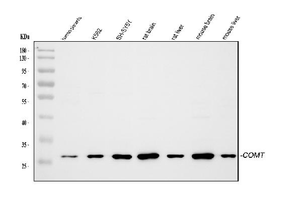
Figure 1. Western blot analysis of COMT using anti-COMT antibody (PA2203). Electrophoresis was performed on a 5-20% SDS-PAGE gel at 70V (Stacking gel) / 90V (Resolving gel) for 2-3 hours. The sample well of each lane was loaded with 30 ug of sample under reducing conditions. Lane 1: human placenta tissue lysates, Lane 2: human K562 whole cell lysates, Lane 3: human SH-SY5Y whole cell lysates, Lane 4: rat brain tissue lysates, Lane 5: rat liver tissue lysates, Lane 6: mouse brain tissue lysates, Lane 7: mouse liver tissue lysates. After electrophoresis, proteins were transferred to a nitrocellulose membrane at 150 mA for 50-90 minutes. Blocked the membrane with 5% non-fat milk/TBS for 1.5 hour at RT. The membrane was incubated with rabbit anti-COMT antigen affinity purified polyclonal antibody (Catalog # PA2203) at 0.5 microg/mL overnight at 4°C, then washed with TBS-0.1%Tween 3 times with 5 minutes each and probed with a goat anti-rabbit IgG-HRP secondary antibody at a dilution of 1:5000 for 1.5 hour at RT. The signal is developed using an Enhanced Chemiluminescent detection (ECL) kit (Catalog # EK1002) with Tanon 5200 system. A specific band was detected for COMT at approximately 28 kDa. The expected band size for COMT is at 30 kDa.
. COMT was detected in a paraffin-embedded section of Human Kidney Cancer tissue. Heat mediated antigen retrieval was performed in EDTA buffer (pH 8.0, epitope retrieval solution). The tissue section was blocked with 10% goat serum. The tissue section was then incubated with 1 microg/ml rabbit anti-COMT Antibody (PA2203) overnight at 4°C. Peroxidase Conjugated Goat Anti-rabbit IgG was used as secondary antibody and incubated for 30 minutes at 37°C. The tissue section was developed using HRP Conjugated Rabbit IgG Super Vision Assay Kit (Catalog # SV0002) with DAB as the chromogen.")
. COMT was detected in a paraffin-embedded section of Human Lung Cancer tissue. Heat mediated antigen retrieval was performed in EDTA buffer (pH 8.0, epitope retrieval solution). The tissue section was blocked with 10% goat serum. The tissue section was then incubated with 1 microg/ml rabbit anti-COMT Antibody (PA2203) overnight at 4°C. Peroxidase Conjugated Goat Anti-rabbit IgG was used as secondary antibody and incubated for 30 minutes at 37°C. The tissue section was developed using HRP Conjugated Rabbit IgG Super Vision Assay Kit (Catalog # SV0002) with DAB as the chromogen.")
Figure 1. Western blot analysis of COMT using anti-COMT antibody (PA2203). Electrophoresis was performed on a 5-20% SDS-PAGE gel at 70V (Stacking gel) / 90V (Resolving gel) for 2-3 hours. The sample well of each lane was loaded with 30 ug of sample under reducing conditions. Lane 1: human placenta tissue lysates, Lane 2: human K562 whole cell lysates, Lane 3: human SH-SY5Y whole cell lysates, Lane 4: rat brain tissue lysates, Lane 5: rat liver tissue lysates, Lane 6: mouse brain tissue lysates, Lane 7: mouse liver tissue lysates. After electrophoresis, proteins were transferred to a nitrocellulose membrane at 150 mA for 50-90 minutes. Blocked the membrane with 5% non-fat milk/TBS for 1.5 hour at RT. The membrane was incubated with rabbit anti-COMT antigen affinity purified polyclonal antibody (Catalog # PA2203) at 0.5 microg/mL overnight at 4°C, then washed with TBS-0.1%Tween 3 times with 5 minutes each and probed with a goat anti-rabbit IgG-HRP secondary antibody at a dilution of 1:5000 for 1.5 hour at RT. The signal is developed using an Enhanced Chemiluminescent detection (ECL) kit (Catalog # EK1002) with Tanon 5200 system. A specific band was detected for COMT at approximately 28 kDa. The expected band size for COMT is at 30 kDa.
Anti-COMT Antibody
PA2203
ApplicationsWestern Blot, ImmunoHistoChemistry
Product group Antibodies
ReactivityHuman, Mouse, Rat
TargetCOMT
Overview
- SupplierBoster Bio
- Product NameAnti-COMT Antibody
- Delivery Days Customer9
- Application Supplier NoteTested Species: In-house tested species with positive results. Predicted Species: Species predicted to be fit for the product based on sequence similarities. By Heat: Boiling the paraffin sections in 10mM citrate buffer, pH6.0, for 20mins is required for the staining of formalin/paraffin sections. Other applications have not been tested. Optimal dilutions should be determined by end users.
- ApplicationsWestern Blot, ImmunoHistoChemistry
- Applications SupplierIHP, WB, IHC
- CertificationResearch Use Only
- ClonalityPolyclonal
- Concentration500 ug/ml
- Gene ID1312
- Target nameCOMT
- Target descriptioncatechol-O-methyltransferase
- Target synonymsHEL-S-98n, catechol O-methyltransferase, epididymis secretory sperm binding protein Li 98n, testicular tissue protein Li 42
- HostRabbit
- IsotypeIgG
- Protein IDP21964
- Protein NameCatechol O-methyltransferase
- Scientific DescriptionBoster Bio Anti-Catechol O-methyltransferase COMT Antibody catalog # PA2203. Tested in IHC, WB applications. This antibody reacts with Human, Mouse, Rat. The brand Picoband indicates this is a premium antibody that guarantees superior quality, high affinity, and strong signals with minimal background in Western blot applications. Only our best-performing antibodies are designated as Picoband, ensuring unmatched performance.
- ReactivityHuman, Mouse, Rat
- Reactivity SupplierHuman
- Storage Instruction-20°C,2°C to 8°C
- UNSPSC12352203
Datasheet
MSDS
Related products
Product group Antibodies
Anti-COMT Antibody144-06200
ApplicationsImmunoFluorescence, Western Blot, ImmunoHistoChemistry
ReactivityHuman, Mouse, Rat
TargetCOMT
- SizePrice

Product group Antibodies
References
COMT antibodyGTX101233
ApplicationsWestern Blot, ELISA, ImmunoHistoChemistry, ImmunoHistoChemistry Paraffin
ReactivityHuman, Mouse
TargetCOMT
- SizePrice
Product group Antibodies
ApplicationsImmunoFluorescence, Western Blot, ImmunoCytoChemistry, ImmunoHistoChemistry
TargetCOMT
- SizePrice
Product group Antibodies
ApplicationsFlow Cytometry, Western Blot, ImmunoCytoChemistry
ReactivityHuman, Rat
TargetCOMT
- SizePrice
Product group Antibodies
COMT AntibodyCSB-PA007149
ApplicationsWestern Blot, ELISA
ReactivityHuman
TargetCOMT
- SizePrice
Product group Antibodies
Anti-COMT AntibodyA101488
ApplicationsWestern Blot, ELISA
ReactivityHuman
- SizePrice
Product group Antibodies

COMT AntibodyLS-C830464
ApplicationsWestern Blot, ELISA, ImmunoHistoChemistry
ReactivityHuman
TargetCOMT
- SizePrice

Product group Antibodies
References

ApplicationsFlow Cytometry, Western Blot, ELISA
ReactivityCanine, Human
TargetCOMT
- SizePrice