

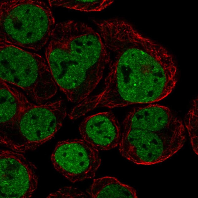
Immunofluorescent staining of human cell line HEL shows localization to nucleoplasm.
Immunofluorescent staining of human cell line HEL shows localization to nucleoplasm.
Anti-CX3CR1 Antibody
HPA030311
ApplicationsImmunoCytoChemistry
Product group Antibodies
ReactivityHuman
TargetCX3CR1
Overview
- SupplierAtlas Antibodies
- Product NameAnti-CX3CR1 Antibody
- Delivery Days Customer4
- ApplicationsImmunoCytoChemistry
- CertificationResearch Use Only
- ClonalityPolyclonal
- ConjugateUnconjugated
- Gene ID1524
- Target nameCX3CR1
- Target descriptionC-X3-C motif chemokine receptor 1
- Target synonymsCCRL1, CMKBRL1, CMKDR1, GPR13, GPRV28, V28, CX3C chemokine receptor 1, C-X3-C CKR-1, CMK-BRL-1, CMK-BRL1, G-protein coupled receptor 13, beta chemokine receptor-like 1, chemokine (C-C) receptor-like 1, chemokine (C-X3-C motif) receptor 1, chemokine (C-X3-C) receptor 1, fractalkine receptor
- HostRabbit
- IsotypeIgG
- Protein IDP49238
- Protein NameCX3C chemokine receptor 1
- Scientific DescriptionRecombinant Protein Epitope Signature Tag (PrEST) antigen sequence
- ReactivityHuman
- Storage Instruction-20°C,2°C to 8°C
- UNSPSC41116161
Datasheet
MSDS
Related products
Product group Antibodies
Anti-CX3CR1 AntibodyA82937
ApplicationsWestern Blot, ELISA
ReactivityHuman
- SizePrice
Product group Antibodies
Anti-CX3CR1 (N-term) Antibody102-22537
ApplicationsWestern Blot
TargetCX3CR1
- SizePrice

Product group Antibodies
Anti-CX3CR1 AntibodyA00280-2-CARRIER-FREE
ApplicationsFlow Cytometry, ELISA, ImmunoHistoChemistry
ReactivityHuman
TargetCX3CR1
- SizePrice
Product group Antibodies

Goat anti-CX3CR1 (aa34-47)EB12515
ApplicationsWestern Blot, ELISA
ReactivityHuman
TargetCX3CR1
- SizePrice
Product group Antibodies
ApplicationsImmunoPrecipitation, Western Blot, ImmunoCytoChemistry, ImmunoHistoChemistry
TargetCX3CR1
- SizePrice

Product group Antibodies
CX3CR1 AntibodyCSB-PA004776
ApplicationsWestern Blot, ELISA
ReactivityHuman, Mouse, Rat
TargetCX3CR1
- SizePrice
Product group Antibodies
References
CX3CR1 Polyclonal AntibodyBS-1728R
ApplicationsFlow Cytometry, Western Blot, ELISA, ImmunoHistoChemistry, ImmunoHistoChemistry Frozen, ImmunoHistoChemistry Paraffin
ReactivityBovine, Canine, Human, Mouse, Rabbit, Rat
TargetCX3CR1
- SizePrice