


Figure 1. IHC analysis of CXCR3 using anti-CXCR3 antibody (PB9079). CXCR3 was detected in a paraffin-embedded section of rat kidney tissue. Heat mediated antigen retrieval was performed in EDTA buffer (pH 8.0, epitope retrieval solution). The tissue section was blocked with 10% goat serum. The tissue section was then incubated with 1 microg/ml rabbit anti-CXCR3 Antibody (PB9079) overnight at 4°C. Biotinylated goat anti-rabbit IgG was used as secondary antibody and incubated for 30 minutes at 37°C. The tissue section was developed using Strepavidin-Biotin-Complex (SABC) (Catalog # SA1022) with DAB as the chromogen.
. CXCR3 was detected in a paraffin-embedded section of human tonsil tissue. Heat mediated antigen retrieval was performed in EDTA buffer (pH 8.0, epitope retrieval solution). The tissue section was blocked with 10% goat serum. The tissue section was then incubated with 1 microg/ml rabbit anti-CXCR3 Antibody (PB9079) overnight at 4°C. Biotinylated goat anti-rabbit IgG was used as secondary antibody and incubated for 30 minutes at 37°C. The tissue section was developed using Strepavidin-Biotin-Complex (SABC) (Catalog # SA1022) with DAB as the chromogen.")
. CXCR3 was detected in a paraffin-embedded section of human lung cancer tissue. Heat mediated antigen retrieval was performed in EDTA buffer (pH 8.0, epitope retrieval solution). The tissue section was blocked with 10% goat serum. The tissue section was then incubated with 1 microg/ml rabbit anti-CXCR3 Antibody (PB9079) overnight at 4°C. Biotinylated goat anti-rabbit IgG was used as secondary antibody and incubated for 30 minutes at 37°C. The tissue section was developed using Strepavidin-Biotin-Complex (SABC) (Catalog # SA1022) with DAB as the chromogen.")
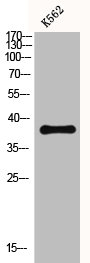
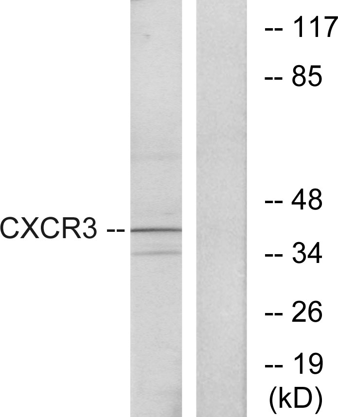
. Electrophoresis was performed on a 5-20% SDS-PAGE gel at 70V (Stacking gel) / 90V (Resolving gel) for 2-3 hours. The sample well of each lane was loaded with 30 ug of sample under reducing conditions. Lane 1: human Colo320 whole cell lysates, Lane 2: human SGC whole cell lysates. After electrophoresis, proteins were transferred to a nitrocellulose membrane at 150 mA for 50-90 minutes. Blocked the membrane with 5% non-fat milk/TBS for 1.5 hour at RT. The membrane was incubated with rabbit anti-CXCR3 antigen affinity purified polyclonal antibody (Catalog # PB9079) at 0.5 microg/mL overnight at 4°C, then washed with TBS-0.1%Tween 3 times with 5 minutes each and probed with a goat anti-rabbit IgG-HRP secondary antibody at a dilution of 1:5000 for 1.5 hour at RT. The signal is developed using an Enhanced Chemiluminescent detection (ECL) kit (Catalog # EK1002) with Tanon 5200 system. A specific band was detected for CXCR3 at approximately 41 kDa. The expected band size for CXCR3 is at 41 kDa.")
Figure 1. IHC analysis of CXCR3 using anti-CXCR3 antibody (PB9079). CXCR3 was detected in a paraffin-embedded section of rat kidney tissue. Heat mediated antigen retrieval was performed in EDTA buffer (pH 8.0, epitope retrieval solution). The tissue section was blocked with 10% goat serum. The tissue section was then incubated with 1 microg/ml rabbit anti-CXCR3 Antibody (PB9079) overnight at 4°C. Biotinylated goat anti-rabbit IgG was used as secondary antibody and incubated for 30 minutes at 37°C. The tissue section was developed using Strepavidin-Biotin-Complex (SABC) (Catalog # SA1022) with DAB as the chromogen.
Anti-CXCR3 Antibody Picoband(r)
PB9079-CARRIER-FREE
ApplicationsWestern Blot, ImmunoHistoChemistry
Product group Antibodies
ReactivityHuman, Rat
TargetCXCR3
Overview
- SupplierBoster Bio
- Product NameAnti-CXCR3 Antibody Picoband(r)
- Delivery Days Customer9
- Application Supplier NoteWB: The detection limit for CXCR3 is approximately 0.25ng/lane under reducing conditions. Tested Species: In-house tested species with positive results. By Heat: Boiling the paraffin sections in 10mM citrate buffer, pH6.0, for 20mins is required for the staining of formalin/paraffin sections. Other applications have not been tested. Optimal dilutions should be determined by end users.
- ApplicationsWestern Blot, ImmunoHistoChemistry
- CertificationResearch Use Only
- ClonalityPolyclonal
- Concentration500 ug/ml
- Gene ID2833
- Target nameCXCR3
- Target descriptionC-X-C motif chemokine receptor 3
- Target synonymsCD182, CD183, CKR-L2, CMKAR3, GPR9, IP10-R, Mig-R, MigR, C-X-C chemokine receptor type 3, G protein-coupled receptor 9, IP-10 receptor, Mig receptor, chemokine (C-X-C motif) receptor 3, chemokine receptor 3, interferon-inducible protein 10 receptor
- HostRabbit
- IsotypeIgG
- Protein IDP49682
- Protein NameC-X-C chemokine receptor type 3
- Scientific DescriptionBoster Bio Anti-CXCR3 Antibody Picoband® catalog # PB9079. Tested in IHC, WB applications. This antibody reacts with Human, Rat. The brand Picoband indicates this is a premium antibody that guarantees superior quality, high affinity, and strong signals with minimal background in Western blot applications. Only our best-performing antibodies are designated as Picoband, ensuring unmatched performance.
- ReactivityHuman, Rat
- Storage Instruction-20°C,2°C to 8°C
- UNSPSC12352203
Related products
Product group Antibodies
CXCR3 AntibodyCSB-PA001843
ApplicationsWestern Blot, ELISA
ReactivityHuman
TargetCXCR3
- SizePrice
Product group Antibodies
Anti-CXCR3 AntibodyA101125
ApplicationsWestern Blot, ELISA
ReactivityHuman
- SizePrice
Product group Antibodies
Anti-Human CD183 Antibody, APC136-25078
ApplicationsFlow Cytometry
ReactivityHuman
TargetCXCR3
- SizePrice
Product group Antibodies

Anti-CXCR3 [9C5]Ab02335-1.1
ApplicationsFlow Cytometry, ELISA
ReactivityHuman, Mouse, Primate
TargetCXCR3
- SizePrice
Product group Antibodies

CXCR3 Antibody (clone CXCR3-173)LS-C772820
ApplicationsFlow Cytometry
ReactivityMouse
TargetCXCR3
- SizePrice
Product group Antibodies

Goat anti-CXCR3 / GPR9EB07201
ApplicationsFlow Cytometry, ImmunoFluorescence, ELISA, ImmunoHistoChemistry
ReactivityHuman, Mouse, Rat
TargetCXCR3
- SizePrice

Product group Antibodies
Anti-CXCR3 AntibodyHPA045942
ApplicationsImmunoHistoChemistry
ReactivityHuman
TargetCXCR3
- SizePrice
Product group Antibodies
CXCR3 Polyclonal AntibodyBS-0341R
ApplicationsFlow Cytometry, ImmunoFluorescence, Western Blot, ELISA, ImmunoCytoChemistry, ImmunoHistoChemistry, ImmunoHistoChemistry Frozen, ImmunoHistoChemistry Paraffin
ReactivityBovine, Canine, Guinea Pig, Human, Mouse, Porcine, Rabbit, Rat
TargetCXCR3
- SizePrice
![CXCR3 antibody [HL3089] detects CXCR3 protein by immunohistochemical analysis. Sample: Paraffin-embedded mouse thymus gland. CXCR3 stained by CXCR3 antibody [HL3089] (GTX640535) diluted at 1:100. Antigen Retrieval: Citrate buffer, pH 6.0, 15 min](https://www.genetex.com/upload/website/prouct_img/normal/GTX640535/GTX640535_T-45453_20240802_IHC-P_M_24081300_546.webp)
Product group Antibodies
CXCR3 antibody [HL3089]GTX640535
ApplicationsImmunoHistoChemistry, ImmunoHistoChemistry Paraffin
ReactivityHuman, Mouse
TargetCXCR3
- SizePrice