

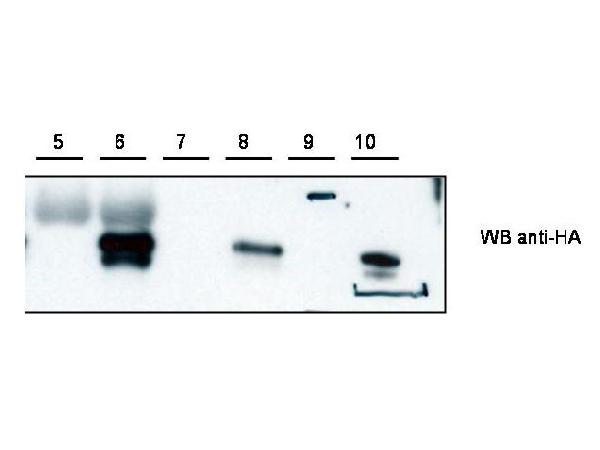
Figure 1. Western blot analysis of CYTIP using anti-CYTIP antibody (A09710). Electrophoresis was performed on a 5-20% SDS-PAGE gel at 70V (Stacking gel) / 90V (Resolving gel) for 2-3 hours. The sample well of each lane was loaded with 50ug of sample under reducing conditions. After Electrophoresis, proteins were transferred to a Nitrocellulose membrane at 150mA for 50-90 minutes. Blocked the membrane with 5% Non-fat Milk/ TBS for 1.5 hour at RT. The membrane was incubated with rabbit anti-CYTIP antigen affinity purified polyclonal antibody (Catalog # A09710) at 0.5 ug/mL overnight at 4°C, then washed with TBS-0.1%Tween 3 times with 5 minutes each and probed with a goat anti-Rabbit IgG-HRP secondary antibody at a dilution of 1:10000 for 1.5 hour at RT. The signal is developed using an Enhanced Chemiluminescent detection (ECL) kit (Catalog # SA1022) with Tanon 5200 system. A specific band was detected for CYTIP.
Figure 1. Western blot analysis of CYTIP using anti-CYTIP antibody (A09710). Electrophoresis was performed on a 5-20% SDS-PAGE gel at 70V (Stacking gel) / 90V (Resolving gel) for 2-3 hours. The sample well of each lane was loaded with 50ug of sample under reducing conditions. After Electrophoresis, proteins were transferred to a Nitrocellulose membrane at 150mA for 50-90 minutes. Blocked the membrane with 5% Non-fat Milk/ TBS for 1.5 hour at RT. The membrane was incubated with rabbit anti-CYTIP antigen affinity purified polyclonal antibody (Catalog # A09710) at 0.5 ug/mL overnight at 4°C, then washed with TBS-0.1%Tween 3 times with 5 minutes each and probed with a goat anti-Rabbit IgG-HRP secondary antibody at a dilution of 1:10000 for 1.5 hour at RT. The signal is developed using an Enhanced Chemiluminescent detection (ECL) kit (Catalog # SA1022) with Tanon 5200 system. A specific band was detected for CYTIP.
Anti-Cybr CYTIP Antibody
A09710
ApplicationsImmunoPrecipitation, Western Blot, ELISA
Product group Antibodies
ReactivityHuman, Mouse, Rat
TargetCytip
Overview
- SupplierBoster Bio
- Product NameAnti-Cybr CYTIP Antibody
- Delivery Days Customer9
- ApplicationsImmunoPrecipitation, Western Blot, ELISA
- CertificationResearch Use Only
- ClonalityPolyclonal
- Concentration85 mg/ml
- Gene ID227929
- Target nameCytip
- Target descriptioncytohesin 1 interacting protein
- Target synonymsA130053M09Rik, Cbp, Cybr, Pscdbp, cytohesin-interacting protein, cbp HE, cytohesin binding protein, cytohesin-binding protein HE, pleckstrin homology, Sec7 and coiled-coil domains, binding protein, tamalin
- HostRabbit
- Protein IDQ91VY6
- Protein NameCytohesin-interacting protein
- Scientific DescriptionBoster Bio Anti-Cybr CYTIP Antibody (Catalog # A09710). Tested in ELISA, IP, WB applications. This antibody reacts with Human, Mouse, Rat.
- ReactivityHuman, Mouse, Rat
- Storage Instruction-20°C,2°C to 8°C
- UNSPSC12352203
Related products
Product group Antibodies
PSCDBP antibodyGTX48718
ApplicationsWestern Blot, ELISA
ReactivityMouse
TargetCytip
- SizePrice