


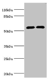
Figure 1. Western blot analysis of Cytochrome P450 2E1/CYP2E1 using anti-Cytochrome P450 2E1/CYP2E1 antibody (M00672-2). Electrophoresis was performed on a 5-20% SDS-PAGE gel at 70V (Stacking gel) / 90V (Resolving gel) for 2-3 hours. The sample well of each lane was loaded with 30 ug of sample under reducing conditions. Lane 1: human HCCP tissue lysates, Lane 2: rat liver tissue lysates, Lane 3: mouse liver tissue lysates. After electrophoresis, proteins were transferred to a nitrocellulose membrane at 150 mA for 50-90 minutes. Blocked the membrane with 5% non-fat milk/TBS for 1.5 hour at RT. The membrane was incubated with mouse anti-Cytochrome P450 2E1/CYP2E1 antigen affinity purified monoclonal antibody (Catalog # M00672-2) at 0.5 microg/mL overnight at 4°C, then washed with TBS-0.1%Tween 3 times with 5 minutes each and probed with a goat anti-mouse IgG-HRP secondary antibody at a dilution of 1:10000 for 1.5 hour at RT. The signal is developed using an Enhanced Chemiluminescent detection (ECL) kit (Catalog # EK1001) with Tanon 5200 system. A specific band was detected for Cytochrome P450 2E1/CYP2E1 at approximately 56 kDa. The expected band size for Cytochrome P450 2E1/CYP2E1 is at 56 kDa.
Figure 1. Western blot analysis of Cytochrome P450 2E1/CYP2E1 using anti-Cytochrome P450 2E1/CYP2E1 antibody (M00672-2). Electrophoresis was performed on a 5-20% SDS-PAGE gel at 70V (Stacking gel) / 90V (Resolving gel) for 2-3 hours. The sample well of each lane was loaded with 30 ug of sample under reducing conditions. Lane 1: human HCCP tissue lysates, Lane 2: rat liver tissue lysates, Lane 3: mouse liver tissue lysates. After electrophoresis, proteins were transferred to a nitrocellulose membrane at 150 mA for 50-90 minutes. Blocked the membrane with 5% non-fat milk/TBS for 1.5 hour at RT. The membrane was incubated with mouse anti-Cytochrome P450 2E1/CYP2E1 antigen affinity purified monoclonal antibody (Catalog # M00672-2) at 0.5 microg/mL overnight at 4°C, then washed with TBS-0.1%Tween 3 times with 5 minutes each and probed with a goat anti-mouse IgG-HRP secondary antibody at a dilution of 1:10000 for 1.5 hour at RT. The signal is developed using an Enhanced Chemiluminescent detection (ECL) kit (Catalog # EK1001) with Tanon 5200 system. A specific band was detected for Cytochrome P450 2E1/CYP2E1 at approximately 56 kDa. The expected band size for Cytochrome P450 2E1/CYP2E1 is at 56 kDa.
Anti-Cytochrome P450 2E1/CYP2E1 Antibody Picoband(r) (monoclonal, 2C7G1)
M00672-2-CARRIER-FREE
ApplicationsWestern Blot
Product group Antibodies
ReactivityHuman, Mouse, Rat
TargetCYP2E1
Overview
- SupplierBoster Bio
- Product NameAnti-Cytochrome P450 2E1/CYP2E1 Antibody Picoband(r) (monoclonal, 2C7G1)
- Delivery Days Customer9
- ApplicationsWestern Blot
- CertificationResearch Use Only
- ClonalityMonoclonal
- Clone ID2C7G1
- Concentration500 ug/ml
- Gene ID1571
- Target nameCYP2E1
- Target descriptioncytochrome P450 family 2 subfamily E member 1
- Target synonymsCPE1, CYP2E, P450-J, P450C2E, cytochrome P450 2E1, 4-nitrophenol 2-hydroxylase, CYPIIE1, cytochrome P450, family 2, subfamily E, polypeptide 1, cytochrome P450, subfamily IIE (ethanol-inducible), polypeptide 1, cytochrome P450-J, flavoprotein-linked monooxygenase, microsomal monooxygenase, xenobiotic monooxygenase
- HostMouse
- IsotypeIgG2b
- Protein IDP05181
- Protein NameCytochrome P450 2E1
- Scientific DescriptionBoster Bio Anti-Cytochrome P450 2E1/CYP2E1 Antibody Picoband® (monoclonal, 2C7G1) catalog # M00672-2. Tested in WB applications. This antibody reacts with Human, Mouse, Rat. The brand Picoband indicates this is a premium antibody that guarantees superior quality, high affinity, and strong signals with minimal background in Western blot applications. Only our best-performing antibodies are designated as Picoband, ensuring unmatched performance.
- ReactivityHuman, Mouse, Rat
- Storage Instruction-20°C,2°C to 8°C
- UNSPSC12352203
Related products
Product group Antibodies
Anti-CYP2E1 Antibody144-61331
ApplicationsImmunoFluorescence, Western Blot, ImmunoHistoChemistry
ReactivityHuman, Mouse, Rat
TargetCYP2E1
- SizePrice
Product group Antibodies
Cyp2E1 Polyclonal AntibodyCAC07078
ApplicationsWestern Blot, ELISA, ImmunoHistoChemistry
ReactivityMouse
TargetCYP2E1
- SizePrice
Product group Antibodies
CYP2E1 AntibodyCSB-PA006425EA01HU
ApplicationsWestern Blot, ELISA, ImmunoHistoChemistry
ReactivityHuman, Mouse
TargetCYP2E1
- SizePrice
Product group Antibodies
ApplicationsImmunoFluorescence, ELISA, ImmunoHistoChemistry
ReactivityHuman, Mouse, Rat
- SizePrice
Product group Antibodies
References
CYP2E1 antibodyGTX32546
ApplicationsImmunoFluorescence, Western Blot, ImmunoCytoChemistry, ImmunoHistoChemistry, ImmunoHistoChemistry Paraffin
ReactivityHuman, Mouse, Rat
TargetCYP2E1
- SizePrice
Product group Antibodies
References
CYP2E1 Polyclonal AntibodyBS-4562R
ApplicationsImmunoFluorescence, Western Blot, ELISA, ImmunoCytoChemistry, ImmunoHistoChemistry, ImmunoHistoChemistry Frozen, ImmunoHistoChemistry Paraffin
ReactivityBovine, Canine, Equine, Human, Mouse, Porcine, Rabbit, Rat, Sheep
TargetCYP2E1
- SizePrice

Product group Antibodies
Anti-CYP2E1 AntibodyHPA009128
ApplicationsWestern Blot, ImmunoHistoChemistry
ReactivityHuman, Mouse, Rat
TargetCYP2E1
- SizePrice
Product group Antibodies

Goat anti-CYP2E1 AntibodyEB10355
ApplicationsWestern Blot, ELISA, ImmunoHistoChemistry
ReactivityHuman
TargetCYP2E1
- SizePrice