


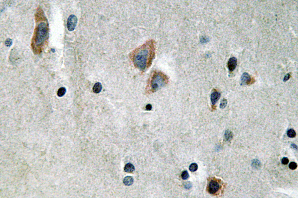
Figure 4. IHC analysis of Cytokeratin 14 using anti-Cytokeratin 14 antibody (A01432). Cytokeratin 14 was detected in paraffin-embedded section of human oesophagus squama cancer tissues. Heat mediated antigen retrieval was performed in citrate buffer (pH6, epitope retrieval solution) for 20 mins. The tissue section was blocked with 10% goat serum. The tissue section was then incubated with 1microg/ml rabbit anti-Cytokeratin 14 Antibody (A01432) overnight at 4°C. Biotinylated goat anti-rabbit IgG was used as secondary antibody and incubated for 30 minutes at 37°C. The tissue section was developed using Strepavidin-Biotin-Complex (SABC)(Catalog # SA1022) with DAB as the chromogen.
. Cytokeratin 14 was detected in paraffin-embedded section of human tonsil tissues. Heat mediated antigen retrieval was performed in citrate buffer (pH6, epitope retrieval solution) for 20 mins. The tissue section was blocked with 10% goat serum. The tissue section was then incubated with 1microg/ml rabbit anti-Cytokeratin 14 Antibody (A01432) overnight at 4°C. Biotinylated goat anti-rabbit IgG was used as secondary antibody and incubated for 30 minutes at 37°C. The tissue section was developed using Strepavidin-Biotin-Complex (SABC)(Catalog # SA1022) with DAB as the chromogen.")
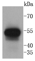
. Electrophoresis was performed on a 5-20% SDS-PAGE gel at 70V (Stacking gel) / 90V (Resolving gel) for 2-3 hours. The sample well of each lane was loaded with 50ug of sample under reducing conditions. lane 1: rat brain tissue lysates, lane 2: NIH3T3 whole cell lysates, lane 3: HEPG2 whole cell lysates After Electrophoresis, proteins were transferred to a Nitrocellulose membrane at 150mA for 50-90 minutes. Blocked the membrane with 5% Non-fat Milk/ TBS for 1.5 hour at RT. The membrane was incubated with rabbit anti-Cytokeratin 14 antigen affinity purified polyclonal antibody (Catalog # A01432) at 0.5 microg/mL overnight at 4°C, then washed with TBS-0.1%Tween 3 times with 5 minutes each and probed with a goat anti-rabbit IgG-HRP secondary antibody at a dilution of 1:10000 for 1.5 hour at RT. The signal is developed using an Enhanced Chemiluminescent detection (ECL) kit (Catalog # EK1002) with Tanon 5200 system. A specific band was detected for Cytokeratin 14 at approximately 60KD. The expected band size for Cytokeratin 14 is at 51KD.")
. Cytokeratin 14 was detected in paraffin-embedded section of human oesophagus squama cancer tissues. Heat mediated antigen retrieval was performed in citrate buffer (pH6, epitope retrieval solution) for 20 mins. The tissue section was blocked with 10% goat serum. The tissue section was then incubated with 1microg/ml rabbit anti-Cytokeratin 14 Antibody (A01432) overnight at 4°C. Biotinylated goat anti-rabbit IgG was used as secondary antibody and incubated for 30 minutes at 37°C. The tissue section was developed using Strepavidin-Biotin-Complex (SABC)(Catalog # SA1022) with DAB as the chromogen.")
. Overlay histogram showing SiHa cells stained with A01432 (Blue line). To facilitate intracellular staining, cells were fixed with 4% paraformaldehyde and permeabilized with permeabilization buffer. The cells were blocked with 10% normal goat serum. And then incubated with rabbit anti-Cytokeratin 14 Antibody (A01432,1microg/1x106 cells) for 30 min at 20°C. DyLight488 conjugated goat anti-rabbit IgG (BA1127, 5-10microg/1x106 cells) was used as secondary antibody for 30 minutes at 20°C. Isotype control antibody (Green line) was rabbit IgG (1microg/1x106) used under the same conditions. Unlabelled sample without incubation with primary antibody and secondary antibody (Red line) was used as a blank control.")
Figure 4. IHC analysis of Cytokeratin 14 using anti-Cytokeratin 14 antibody (A01432). Cytokeratin 14 was detected in paraffin-embedded section of human oesophagus squama cancer tissues. Heat mediated antigen retrieval was performed in citrate buffer (pH6, epitope retrieval solution) for 20 mins. The tissue section was blocked with 10% goat serum. The tissue section was then incubated with 1microg/ml rabbit anti-Cytokeratin 14 Antibody (A01432) overnight at 4°C. Biotinylated goat anti-rabbit IgG was used as secondary antibody and incubated for 30 minutes at 37°C. The tissue section was developed using Strepavidin-Biotin-Complex (SABC)(Catalog # SA1022) with DAB as the chromogen.
Anti-Cytokeratin 14/KRT14 Antibody Picoband(r)
A01432-CARRIER-FREE
ApplicationsFlow Cytometry, Western Blot, ImmunoCytoChemistry, ImmunoHistoChemistry, ImmunoHistoChemistry Frozen
Product group Antibodies
ReactivityHuman, Mouse, Rat
TargetKRT14
Overview
- SupplierBoster Bio
- Product NameAnti-Cytokeratin 14/KRT14 Antibody Picoband(r)
- Delivery Days Customer9
- Application Supplier NoteTested Species: In-house tested species with positive results. By Heat: Boiling the paraffin sections in 10mM citrate buffer, pH6.0, for 20mins is required for the staining of formalin/paraffin sections. Other applications have not been tested. Optimal dilutions should be determined by end users.
- ApplicationsFlow Cytometry, Western Blot, ImmunoCytoChemistry, ImmunoHistoChemistry, ImmunoHistoChemistry Frozen
- CertificationResearch Use Only
- ClonalityPolyclonal
- Concentration500 ug/ml
- Gene ID3861
- Target nameKRT14
- Target descriptionkeratin 14
- Target synonymsCK14, EBS1, EBS1A, EBS1B, EBS1C, EBS1D, EBS3, EBS4, K14, NFJ, keratin, type I cytoskeletal 14, cytokeratin 14, keratin 14, type I
- HostRabbit
- IsotypeIgG
- Protein IDP02533
- Protein NameKeratin, type I cytoskeletal 14
- Scientific DescriptionBoster Bio Anti-Cytokeratin 14/KRT14 Antibody Picoband® catalog # A01432. Tested in Flow Cytometry, IHC, IHC-F, ICC, WB applications. This antibody reacts with Human, Mouse, Rat. The brand Picoband indicates this is a premium antibody that guarantees superior quality, high affinity, and strong signals with minimal background in Western blot applications. Only our best-performing antibodies are designated as Picoband, ensuring unmatched performance.
- ReactivityHuman, Mouse, Rat
- Storage Instruction-20°C,2°C to 8°C
- UNSPSC12352203
Related products
Product group Antibodies
ApplicationsELISA, ImmunoHistoChemistry
ReactivityHuman, Mouse, Rat
- SizePrice
Product group Antibodies

Anti-Cytokeratin 14 [RCK107]AB03338-1.1
ApplicationsFlow Cytometry, Western Blot, ImmunoHistoChemistry
ReactivityCanine, Human, Porcine, Rat
TargetKRT14
- SizePrice
Product group Antibodies
Anti-Cytokeratin 14 Antibody118-10011
ApplicationsELISA, ImmunoHistoChemistry
ReactivityHuman
- SizePrice
Product group Antibodies
Anti-KRT14 AntibodyAMAB91968
ApplicationsWestern Blot, ImmunoCytoChemistry, ImmunoHistoChemistry
ReactivityHuman
TargetKRT14
- SizePrice
Product group Antibodies

KRT14 / CK14 / Cytokeratin 14 AntibodyLS-C835086
ApplicationsImmunoHistoChemistry
ReactivityHuman
TargetKRT14
- SizePrice
Product group Antibodies
Cytokeratin 13 Recombinant AntibodyBSM-52053R
ApplicationsImmunoFluorescence, Western Blot, ImmunoCytoChemistry, ImmunoHistoChemistry, ImmunoHistoChemistry Frozen, ImmunoHistoChemistry Paraffin
ReactivityHuman, Mouse
TargetKRT14
- SizePrice
Product group Antibodies

Cytokeratin 14CK514
ApplicationsImmunoHistoChemistry, ImmunoHistoChemistry Frozen, ImmunoHistoChemistry Paraffin
ReactivityHuman
TargetKRT14
- SizePrice
Product group Antibodies
ApplicationsImmunoPrecipitation, Western Blot, ImmunoCytoChemistry, ImmunoHistoChemistry
ReactivityRat
TargetKRT14
- SizePrice
Product group Antibodies
KRT14 AntibodyCSB-PA005038
ApplicationsWestern Blot, ELISA
ReactivityHuman, Mouse, Rat
TargetKRT14
- SizePrice