

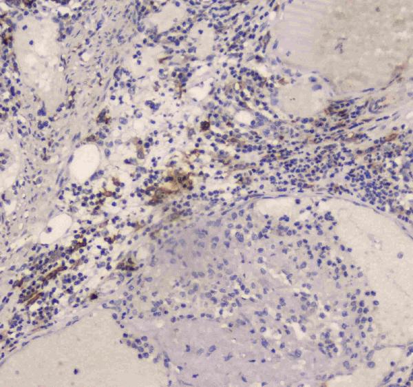
Figure 2. IHC analysis of DC-SIGN using anti-DC-SIGN antibody (A01025-2). DC-SIGN was detected in paraffin-embedded section of human intestinal cancer tissue. Heat mediated antigen retrieval was performed in citrate buffer (pH6, epitope retrieval solution) for 20 mins. The tissue section was blocked with 10% goat serum. The tissue section was then incubated with 1microg/ml rabbit anti-DC-SIGN Antibody (A01025-2) overnight at 4°C. Biotinylated goat anti-rabbit IgG was used as secondary antibody and incubated for 30 minutes at 37°C. The tissue section was developed using Strepavidin-Biotin-Complex (SABC)(Catalog # SA1022) with DAB as the chromogen.
. DC-SIGN was detected in paraffin-embedded section of human placenta tissue. Heat mediated antigen retrieval was performed in citrate buffer (pH6, epitope retrieval solution) for 20 mins. The tissue section was blocked with 10% goat serum. The tissue section was then incubated with 1microg/ml rabbit anti-DC-SIGN Antibody (A01025-2) overnight at 4°C. Biotinylated goat anti-rabbit IgG was used as secondary antibody and incubated for 30 minutes at 37°C. The tissue section was developed using Strepavidin-Biotin-Complex (SABC)(Catalog # SA1022) with DAB as the chromogen.")
. Overlay histogram showing THP-1 cells stained with A01025-2 (Blue line).The cells were blocked with 10% normal goat serum. And then incubated with rabbit anti-DC-SIGN Antibody (A01025-2,1microg/1x106 cells) for 30 min at 20°C. DyLight®488 conjugated goat anti-rabbit IgG (BA1127, 5-10microg/1x106 cells) was used as secondary antibody for 30 minutes at 20°C. Isotype control antibody (Green line) was rabbit IgG (1microg/1x106) used under the same conditions. Unlabelled sample (Red line) was also used as a control.")
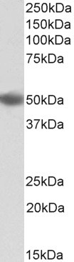
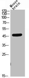
. Electrophoresis was performed on a 5-20% SDS-PAGE gel at 70V (Stacking gel) / 90V (Resolving gel) for 2-3 hours. The sample well of each lane was loaded with 30 ug of sample under reducing conditions. Lane 1: human HepG2 whole cell lysates. After electrophoresis, proteins were transferred to a nitrocellulose membrane at 150 mA for 50-90 minutes. Blocked the membrane with 5% non-fat milk/TBS for 1.5 hour at RT. The membrane was incubated with rabbit anti-DC-SIGN antigen affinity purified polyclonal antibody (Catalog # A01025-2) at 0.5 microg/mL overnight at 4°C, then washed with TBS-0.1%Tween 3 times with 5 minutes each and probed with a goat anti-rabbit IgG-HRP secondary antibody at a dilution of 1:5000 for 1.5 hour at RT. The signal is developed using an Enhanced Chemiluminescent detection (ECL) kit (Catalog # EK1002) with Tanon 5200 system. A specific band was detected for DC-SIGN at approximately 46 kDa. The expected band size for DC-SIGN is at 46 kDa.")
Figure 2. IHC analysis of DC-SIGN using anti-DC-SIGN antibody (A01025-2). DC-SIGN was detected in paraffin-embedded section of human intestinal cancer tissue. Heat mediated antigen retrieval was performed in citrate buffer (pH6, epitope retrieval solution) for 20 mins. The tissue section was blocked with 10% goat serum. The tissue section was then incubated with 1microg/ml rabbit anti-DC-SIGN Antibody (A01025-2) overnight at 4°C. Biotinylated goat anti-rabbit IgG was used as secondary antibody and incubated for 30 minutes at 37°C. The tissue section was developed using Strepavidin-Biotin-Complex (SABC)(Catalog # SA1022) with DAB as the chromogen.
Anti-DC-SIGN/CD209 Antibody Picoband(r)
A01025-2-CARRIER-FREE
ApplicationsFlow Cytometry, Western Blot, ImmunoHistoChemistry
Product group Antibodies
ReactivityHuman
TargetCD209
Overview
- SupplierBoster Bio
- Product NameAnti-DC-SIGN/CD209 Antibody Picoband(r)
- Delivery Days Customer9
- ApplicationsFlow Cytometry, Western Blot, ImmunoHistoChemistry
- CertificationResearch Use Only
- ClonalityPolyclonal
- Concentration500 ug/ml
- Gene ID30835
- Target nameCD209
- Target descriptionCD209 molecule
- Target synonymsCDSIGN, CLEC4L, DC-SIGN, DC-SIGN1, hDC-SIGN, CD209 antigen, C-type lectin domain family 4 member L, HIV gpl20-binding protein, dendritic cell-specific ICAM-3-grabbing non-integrin 1, dendritic cell-specific intercellular adhesion molecule-3-grabbing non-integrin, dendritic cell-specific intracellular adhesion molecules (ICAM)-3 grabbing non-integrin
- HostRabbit
- IsotypeIgG
- Protein IDQ9NNX6
- Protein NameCD209 antigen
- Scientific DescriptionBoster Bio Anti-DC-SIGN/CD209 Antibody Picoband® catalog # A01025-2. Tested in Flow Cytometry, IHC, WB applications. This antibody reacts with Human. The brand Picoband indicates this is a premium antibody that guarantees superior quality, high affinity, and strong signals with minimal background in Western blot applications. Only our best-performing antibodies are designated as Picoband, ensuring unmatched performance.
- ReactivityHuman
- Storage Instruction-20°C,2°C to 8°C
- UNSPSC12352203
Related products
Product group Antibodies
Anti-DC-SIGN AntibodyA83044
ApplicationsWestern Blot, ELISA, ImmunoHistoChemistry
ReactivityHuman
- SizePrice
Product group Antibodies
Anti-Mouse/Rat CD209 Antibody144-01466
ApplicationsWestern Blot
ReactivityHuman, Mouse, Rat
TargetCD209
- SizePrice
Product group Antibodies

DC-SIGN AntibodyABX430798
ApplicationsWestern Blot, ELISA, ImmunoHistoChemistry
- SizePrice
Product group Antibodies

ApplicationsFlow Cytometry, ELISA
ReactivityHuman
TargetCD209
- SizePrice
Product group Antibodies
CD209/DC-SIGN Recombinant Antibody, AbBy Fluor-350 ConjugatedBSM-61560R-BF350
ApplicationsWestern Blot
ReactivityHuman
TargetCD209
- SizePrice
Product group Antibodies
CD209 AntibodyCSB-PA006293
ApplicationsWestern Blot, ELISA
ReactivityHuman, Mouse, Rat
TargetCD209
- SizePrice
Product group Antibodies

Goat anti-DC-SIGN / CD209EB12715
ApplicationsWestern Blot, ELISA
ReactivityHuman
TargetCD209
- SizePrice
Product group Antibodies
CD209 Polyclonal AntibodyCAC14924
ApplicationsImmunoFluorescence, Western Blot, ELISA, ImmunoHistoChemistry
ReactivityMouse
TargetCD209
- SizePrice
Product group Antibodies
DC-SIGN antibodyGTX135602
ApplicationsWestern Blot
ReactivityHuman
TargetCD209
- SizePrice