

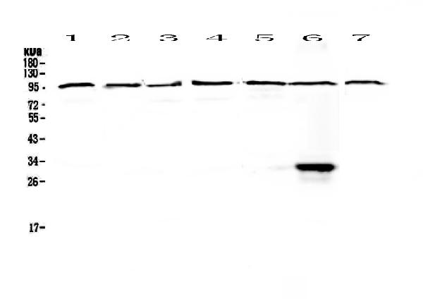
Figure 1. Western blot analysis of DGCR8 using anti-DGCR8 antibody (A00475-1). Electrophoresis was performed on a 5-20% SDS-PAGE gel at 70V (Stacking gel) / 90V (Resolving gel) for 2-3 hours. The sample well of each lane was loaded with 50ug of sample under reducing conditions. Lane 1: human Hela whole cell lysates, Lane 2: human placenta tissue lysates, Lane 3: human COLO-320 whole cell lysates, Lane 4: human HepG2 whole cell lysates, Lane 5: human A549 whole cell lysates, Lane 6: human MCF-7 whole cell lysates, Lane 7: human 22RV1 whole cell lysates. After Electrophoresis, proteins were transferred to a Nitrocellulose membrane at 150mA for 50-90 minutes. Blocked the membrane with 5% Non-fat Milk/ TBS for 1.5 hour at RT. The membrane was incubated with rabbit anti-DGCR8 antigen affinity purified polyclonal antibody (Catalog # A00475-1) at 0.5 microg/mL overnight at 4°C, then washed with TBS-0.1%Tween 3 times with 5 minutes each and probed with a goat anti-rabbit IgG-HRP secondary antibody at a dilution of 1:10000 for 1.5 hour at RT. The signal is developed using an Enhanced Chemiluminescent detection (ECL) kit (Catalog # EK1002) with Tanon 5200 system. A specific band was detected for DGCR8 at approximately 100KD. The expected band size for DGCR8 is at 86KD.
. DGCR8 was detected in paraffin-embedded section of human rectal cancer tissue. Heat mediated antigen retrieval was performed in citrate buffer (pH6, epitope retrieval solution) for 20 mins. The tissue section was blocked with 10% goat serum. The tissue section was then incubated with 1microg/ml rabbit anti-DGCR8 Antibody (A00475-1) overnight at 4°C. Biotinylated goat anti-rabbit IgG was used as secondary antibody and incubated for 30 minutes at 37°C. The tissue section was developed using Strepavidin-Biotin-Complex (SABC)(Catalog # SA1022) with DAB as the chromogen.")
. DGCR8 was detected in paraffin-embedded section of mouse small intestine tissues. Heat mediated antigen retrieval was performed in citrate buffer (pH6, epitope retrieval solution) for 20 mins. The tissue section was blocked with 10% goat serum. The tissue section was then incubated with 1microg/ml rabbit anti-DGCR8 Antibody (A00475-1) overnight at 4°C. Biotinylated goat anti-rabbit IgG was used as secondary antibody and incubated for 30 minutes at 37°C. The tissue section was developed using Strepavidin-Biotin-Complex (SABC)(Catalog # SA1022) with DAB as the chromogen.")
. DGCR8 was detected in immunocytochemical section of A431 cell. Enzyme antigen retrieval was performed using IHC enzyme antigen retrieval reagent (AR0022) for 15 mins. The cells were blocked with 10% goat serum. And then incubated with 2microg/mL rabbit anti-DGCR8 Antibody (A00475-1) overnight at 4°C. DyLight®488 Conjugated Goat Anti-Rabbit IgG (BA1127) was used as secondary antibody at 1:100 dilution and incubated for 30 minutes at 37°C. The section was counterstained with DAPI. Visualize using a fluorescence microscope and filter sets appropriate for the label used.")
Figure 1. Western blot analysis of DGCR8 using anti-DGCR8 antibody (A00475-1). Electrophoresis was performed on a 5-20% SDS-PAGE gel at 70V (Stacking gel) / 90V (Resolving gel) for 2-3 hours. The sample well of each lane was loaded with 50ug of sample under reducing conditions. Lane 1: human Hela whole cell lysates, Lane 2: human placenta tissue lysates, Lane 3: human COLO-320 whole cell lysates, Lane 4: human HepG2 whole cell lysates, Lane 5: human A549 whole cell lysates, Lane 6: human MCF-7 whole cell lysates, Lane 7: human 22RV1 whole cell lysates. After Electrophoresis, proteins were transferred to a Nitrocellulose membrane at 150mA for 50-90 minutes. Blocked the membrane with 5% Non-fat Milk/ TBS for 1.5 hour at RT. The membrane was incubated with rabbit anti-DGCR8 antigen affinity purified polyclonal antibody (Catalog # A00475-1) at 0.5 microg/mL overnight at 4°C, then washed with TBS-0.1%Tween 3 times with 5 minutes each and probed with a goat anti-rabbit IgG-HRP secondary antibody at a dilution of 1:10000 for 1.5 hour at RT. The signal is developed using an Enhanced Chemiluminescent detection (ECL) kit (Catalog # EK1002) with Tanon 5200 system. A specific band was detected for DGCR8 at approximately 100KD. The expected band size for DGCR8 is at 86KD.
Anti-DGCR8 Antibody Picoband(r)
A00475-1-DYLIGHT550
ApplicationsImmunoFluorescence, Western Blot, ELISA, ImmunoCytoChemistry, ImmunoHistoChemistry
Product group Antibodies
ReactivityHuman, Mouse, Rat
TargetDGCR8
Overview
- SupplierBoster Bio
- Product NameAnti-DGCR8 Antibody Picoband(r)
- Delivery Days Customer9
- ApplicationsImmunoFluorescence, Western Blot, ELISA, ImmunoCytoChemistry, ImmunoHistoChemistry
- CertificationResearch Use Only
- ClonalityPolyclonal
- Concentration500 ug/ml
- ConjugateDyLight 550
- Gene ID54487
- Target nameDGCR8
- Target descriptionDGCR8 microprocessor complex subunit
- Target synonymsC22orf12, DGCRK6, Gy1, pasha, microprocessor complex subunit DGCR8, DiGeorge syndrome critical region 8, DiGeorge syndrome critical region gene 8
- HostRabbit
- IsotypeIgG
- Protein IDQ8WYQ5
- Protein NameMicroprocessor complex subunit DGCR8
- Scientific DescriptionBoster Bio Anti-DGCR8 Antibody Picoband® catalog # A00475-1. Tested in ELISA, IF, IHC, ICC, WB applications. This antibody reacts with Human, Mouse, Rat. The brand Picoband indicates this is a premium antibody that guarantees superior quality, high affinity, and strong signals with minimal background in Western blot applications. Only our best-performing antibodies are designated as Picoband, ensuring unmatched performance.
- ReactivityHuman, Mouse, Rat
- Storage Instruction-20°C,2°C to 8°C
- UNSPSC12352203
Related products
Product group Antibodies
DGCR8 Recombinant AntibodyBSM-61214R
ApplicationsImmunoFluorescence, ImmunoPrecipitation, Western Blot, ImmunoCytoChemistry
TargetDGCR8
- SizePrice
Product group Antibodies
Dgcr8 Recombinant AntibodyCAC12132
ApplicationsWestern Blot, ELISA
TargetDGCR8
- SizePrice
Product group Antibodies
Anti-DGCR8 AntibodyA32381
ApplicationsImmunoFluorescence, Western Blot, ImmunoCytoChemistry
ReactivityHuman, Mouse, Rat
- SizePrice
Product group Antibodies
Anti-DGCR8 (Center) Antibody102-27632
ApplicationsFlow Cytometry, Western Blot
TargetDGCR8
- SizePrice
Product group Antibodies

ApplicationsELISA
ReactivityBovine, Human, Mouse, Rat
TargetDGCR8
- SizePrice
Product group Antibodies
References
DGCR8 antibodyGTX130061
ApplicationsImmunoPrecipitation, Western Blot
ReactivityAmphibian, Human
TargetDGCR8
- SizePrice
Product group Antibodies

DGCR8 Antibody (aa1-50)LS-C286892
ApplicationsImmunoHistoChemistry
ReactivityHuman
TargetDGCR8
- SizePrice

Product group Antibodies
Anti-DGCR8 AntibodyHPA076916
ApplicationsImmunoCytoChemistry
ReactivityHuman
TargetDGCR8
- SizePrice