


Immunohistochemical staining of human spleen shows moderate cytoplasmic positivity in sinusoids.
Immunohistochemical staining of human spleen shows moderate cytoplasmic positivity in sinusoids.
Anti-DIAPH1 Antibody
HPA004916
ApplicationsImmunoHistoChemistry
Product group Antibodies
ReactivityHuman
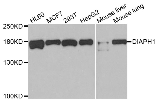
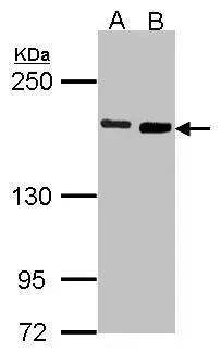
TargetDIAPH1
Overview
- SupplierAtlas Antibodies
- Product NameAnti-DIAPH1 Antibody
- Delivery Days Customer4
- ApplicationsImmunoHistoChemistry
- CertificationResearch Use Only
- ClonalityPolyclonal
- ConjugateUnconjugated
- Gene ID1729
- Target nameDIAPH1
- Target descriptiondiaphanous related formin 1
- Target synonymsDFNA1, DIA1, DRF1, LFHL1, SCBMS, hDIA1, mDia1, protein diaphanous homolog 1, mammalian diaphanous related formin 1
- HostRabbit
- IsotypeIgG
- Protein IDO60610
- Protein NameProtein diaphanous homolog 1
- Scientific DescriptionRecombinant Protein Epitope Signature Tag (PrEST) antigen sequence
- ReactivityHuman
- Storage Instruction-20°C,2°C to 8°C
- UNSPSC41116161
Datasheet
MSDS
Related products
Product group Antibodies
DIAPH1 AntibodyCSB-PA006890GA01HU
ApplicationsImmunoFluorescence, Western Blot, ELISA
ReactivityHuman, Mouse, Rat
TargetDIAPH1
- SizePrice
Product group Antibodies
Anti-DIAPH1 AntibodyA31042
ApplicationsWestern Blot, ImmunoHistoChemistry
ReactivityHuman, Mouse, Rat
- SizePrice

Product group Antibodies
Anti-DIAPH1 Antibody Picoband(r)A02308-3-CARRIER-FREE
ApplicationsFlow Cytometry, ImmunoFluorescence, Western Blot, ELISA, ImmunoCytoChemistry
ReactivityHuman, Monkey
TargetDIAPH1
- SizePrice
Product group Antibodies

DIAPH1 AntibodyLS-C334279
ApplicationsWestern Blot, ImmunoHistoChemistry
ReactivityHuman, Mouse
TargetDIAPH1
- SizePrice
Product group Antibodies
DIAPH1 Recombinant Antibody, AbBy Fluor-555 ConjugatedBSM-61908R-BF555
ApplicationsImmunoFluorescence, Western Blot
ReactivityHuman, Mouse, Rat
TargetDIAPH1
- SizePrice
Product group Antibodies
ApplicationsImmunoPrecipitation, Western Blot, ImmunoCytoChemistry, ImmunoHistoChemistry
TargetDIAPH1
- SizePrice
Product group Antibodies
DIAPH1 antibody [N1N2], N-termGTX102042
ApplicationsImmunoFluorescence, Western Blot, ImmunoCytoChemistry, ImmunoHistoChemistry, ImmunoHistoChemistry Paraffin
ReactivityHuman
TargetDIAPH1
- SizePrice