Bio-Connect
Immunofluorescent staining of human cell line A-431 shows localization to nucleoplasm & cytosol.
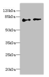
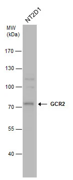
Anti-ECD Antibody

HPA006465
Atlas Antibodies
ApplicationsWestern Blot, ChIP Chromatin ImmunoPrecipitation, ImmunoCytoChemistry
Product group Antibodies
ReactivityHuman
TargetECD
Sign in to order and to see your custom pricing.
Large volume orders?
Order with a bulk request

Overview

  • Supplier
    Atlas Antibodies
  • Product Name
    Anti-ECD
  • Delivery Days Customer
    4
  • Applications
    Western Blot, ChIP Chromatin ImmunoPrecipitation, ImmunoCytoChemistry
  • Certification
    Research Use Only
  • Clonality
    Polyclonal
  • Conjugate
    Unconjugated
  • Gene ID11319
  • Target name
    ECD
  • Target description
    ecdysoneless cell cycle regulator
  • Target synonyms
    ecdysoneless homolog; GCR2; HSGT1; human suppressor of GCR two; protein ecdysoneless homolog; protein SGT1; SGT1; suppressor of GCR2; suppressor of S. cerevisiae gcr2
  • Host
    Rabbit
  • Isotype
    IgG
  • Protein IDO95905
  • Protein Name
    Protein ecdysoneless homolog
  • Scientific Description
    Recombinant Protein Epitope Signature Tag (PrEST) antigen sequence
  • Reactivity
    Human
  • Storage Instruction
    -20°C,2°C to 8°C
  • UNSPSC
    41116161

Related products

Product group Antibodies
Bethyl Laboratories
ApplicationsImmunoPrecipitation, Western Blot
TargetECD
  • SizePrice
Figure 1. Western blot analysis of SGT1/ECD using anti-SGT1/ECD antibody (A00567-1). Electrophoresis was performed on a 5-20% SDS-PAGE gel at 70V (Stacking gel) / 90V (Resolving gel) for 2-3 hours. The sample well of each lane was loaded with 30 ug of sample under reducing conditions. Lane 1: mouse Ana-1 whole cell lysates, Lane 2: human HL-60 whole cell lysates, Lane 3: human U20S whole cell lysates, Lane 4: human U-87MG whole cell lysates, Lane 5: human K562 whole cell lysates, Lane 6: human HepG2 whole cell lysates. After electrophoresis, proteins were transferred to a nitrocellulose membrane at 150 mA for 50-90 minutes. Blocked the membrane with 5% non-fat milk/TBS for 1.5 hour at RT. The membrane was incubated with rabbit anti-SGT1/ECD antigen affinity purified polyclonal antibody (Catalog # A00567-1) at 0.5 microg/mL overnight at 4°C, then washed with TBS-0.1%Tween 3 times with 5 minutes each and probed with a goat anti-rabbit IgG-HRP secondary antibody at a dilution of 1:5000 for 1.5 hour at RT. The signal is developed using an Enhanced Chemiluminescent detection (ECL) kit (Catalog # EK1002) with Tanon 5200 system. A specific band was detected for SGT1/ECD at approximately 73 kDa. The expected band size for SGT1/ECD is at 73 kDa.
Product group Antibodies
Boster Bio
ApplicationsFlow Cytometry, Western Blot, ELISA
TargetECD
  • SizePrice
Product group Antibodies
Biomatik
ApplicationsWestern Blot, ELISA
TargetECD
  • SizePrice
Western blot All lanes: ECD antibody at 6microg/ml Lane 1: Hela whole cell lysate Lane 2: U251 whole cell lysate Secondary Goat polyclonal to rabbit IgG at 1/10000 dilution Predicted band size: 73, 68, 77 kDa Observed band size: 73 kDa
Product group Antibodies
Cusabio
ECD AntibodyCSB-PA007370LA01HU
ApplicationsWestern Blot, ELISA
ReactivityHuman
TargetECD
  • SizePrice
Product group Antibodies
LifeSpan BioSciences
ApplicationsWestern Blot, ELISA
TargetECD
  • SizePrice
Atlas Antibodies
Protein IDO95905
  • SizePrice
Product group Antibodies
GeneTex
ApplicationsWestern Blot
TargetECD
  • SizePrice